

Reliability of diagnostic ultrasound in measuring the multifidus muscle
- PMID: 25878771
- PMCID: PMC4397671
- DOI: 10.1186/s12998-015-0059-6
Reliability of diagnostic ultrasound in measuring the multifidus muscle
Abstract
Background: Ultrasound is frequently used to measure activity in the lumbar multifidus muscle (LMM). However previous reliability studies on diagnostic ultrasound and LMM have included a limited number of subjects and few have used Bland-Altman's Limits of Agreement (LOA). Further one does not know if activity affects the subjects' ability to contract the LMM.
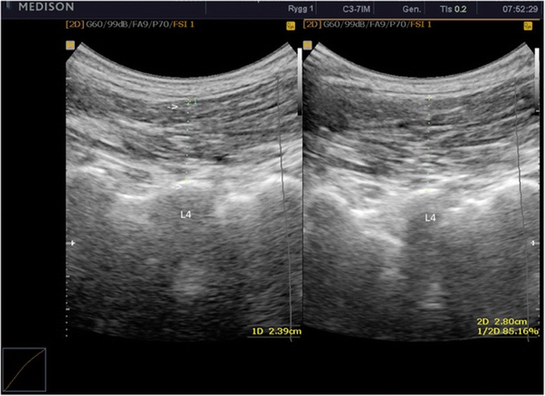
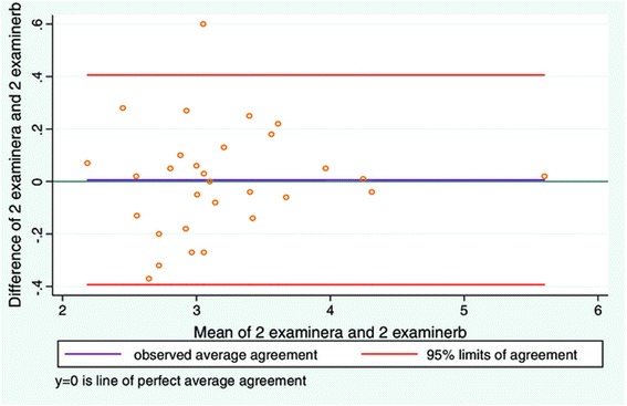
Methods: From January 2012 to December 2012 an inter- and intra-examiner reliability study was carried out in a clinical setting. It consisted of a total of four experiments with 30 subjects in each study. Two experienced examiners performed all measurements. Ultrasound measurements were made of: 1. the LMM in the resting state, 2. during a contracted state, 3. on subsequent days, and, before and after walking. Reliability and agreement was tested for 1. resting LMM, 2. contracted LMM, and 3. thickness change in the LMM. Mean values of three measurements were used for statistical analysis for each spinal level. The intra-class correlation coefficient (ICC) 3.1 and 3.2 was used to test for reliability, and Bland-Altman's LOA method to test for agreement.
Results: All of the studies indicate high levels of reliability, but as the LMM thickness increased (increasing contraction) the agreement between examiners was poorer than for low levels of contraction.
Conclusions: The use of diagnostic ultrasound to measure the LMM seems to be reliable in subjects who have little or no change in thickness of the LMM with contraction.
Keywords: Agreement; Diagnostic ultrasound; Intraclass correlation coefficient; Limits of agreement; Lumbar multifidus; Measurement; Reliability.
Figures


Similar articles
-
Musculoskeletal diagnostic ultrasound imaging for thickness measurement of four principal muscles of the cervical spine -a reliability and agreement study.Chiropr Man Therap. 2017 Jan 4;25:2. doi: 10.1186/s12998-016-0132-9. eCollection 2017. Chiropr Man Therap. 2017. PMID: 28070269 Free PMC article.
-
Ultrasound Imaging Analysis of the Lumbar Multifidus Muscle Echo Intensity: Intra-Rater and Inter-Rater Reliability of a Novice and an Experienced Rater.Medicina (Kaunas). 2021 May 20;57(5):512. doi: 10.3390/medicina57050512. Medicina (Kaunas). 2021. PMID: 34065340 Free PMC article.
-
Reliability of Ultrasound Findings in Patients with Lumbar Multifidus Myofascial Pain Syndrome.Arch Bone Jt Surg. 2023;11(4):248-255. doi: 10.22038/ABJS.2022.63591.3067. Arch Bone Jt Surg. 2023. PMID: 37180289 Free PMC article.
-
Lumbar multifidus muscle ultrasound imaging: Is handheld technology reliable?Musculoskelet Sci Pract. 2023 Jun;65:102771. doi: 10.1016/j.msksp.2023.102771. Epub 2023 Apr 29. Musculoskelet Sci Pract. 2023. PMID: 37182391
-
A Review of the Use of Confidence Intervals for Bland-Altman Limits of Agreement in Optometry and Vision Science.Optom Vis Sci. 2020 Jan;97(1):3-8. doi: 10.1097/OPX.0000000000001465. Optom Vis Sci. 2020. PMID: 31895271 Review.
Cited by
-
Musculoskeletal diagnostic ultrasound imaging for thickness measurement of four principal muscles of the cervical spine -a reliability and agreement study.Chiropr Man Therap. 2017 Jan 4;25:2. doi: 10.1186/s12998-016-0132-9. eCollection 2017. Chiropr Man Therap. 2017. PMID: 28070269 Free PMC article.
-
Estimation of spinopelvic muscles' volumes in young asymptomatic subjects: a quantitative analysis.Surg Radiol Anat. 2017 Apr;39(4):393-403. doi: 10.1007/s00276-016-1742-6. Epub 2016 Sep 16. Surg Radiol Anat. 2017. PMID: 27637762
-
LUMINOUS database: lumbar multifidus muscle segmentation from ultrasound images.BMC Musculoskelet Disord. 2020 Oct 23;21(1):703. doi: 10.1186/s12891-020-03679-3. BMC Musculoskelet Disord. 2020. PMID: 33097024 Free PMC article.
-
Analysis of lumbar spine loading during walking in patients with chronic low back pain and healthy controls: An OpenSim-Based study.Front Bioeng Biotechnol. 2024 May 13;12:1377767. doi: 10.3389/fbioe.2024.1377767. eCollection 2024. Front Bioeng Biotechnol. 2024. PMID: 38817923 Free PMC article.
-
Clinical Efficacy of Multimodal Exercise Telerehabilitation Based on AI for Chronic Nonspecific Low Back Pain: Randomized Controlled Trial.JMIR Mhealth Uhealth. 2025 May 22;13:e56176. doi: 10.2196/56176. JMIR Mhealth Uhealth. 2025. PMID: 40402551 Free PMC article. Clinical Trial.
References
-
- Yoshihara K, Nakayama Y, Fujii N, Aoki T, Ito H. Atrophy of the multifidus muscle in patients with lumbar disk herniation: histochemical and electromyographic study. Orthopedics. 2003;26(5):493–5. - PubMed
LinkOut - more resources
Full Text Sources
Other Literature Sources
Medical
