

A rare case of coronary artery fistula presented with acute myocardial infarction
- PMID: 25878968
- PMCID: PMC4394573
- DOI: 10.4103/2231-0770.154200
A rare case of coronary artery fistula presented with acute myocardial infarction
Abstract
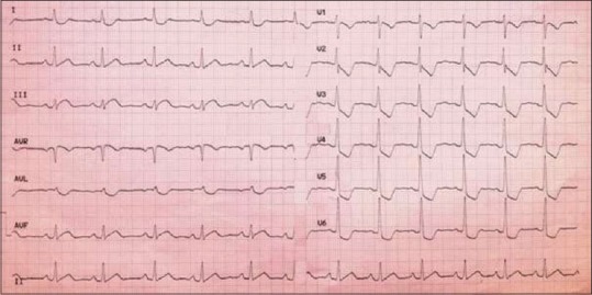
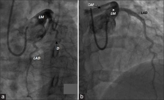
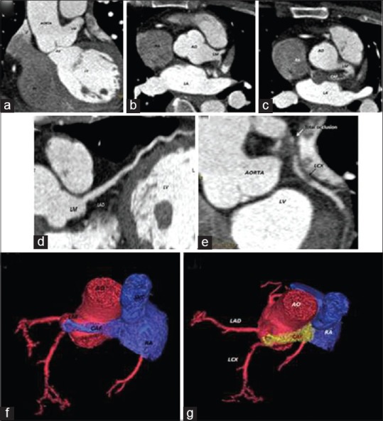
Coronary artery fistulas (CAFs): Are anomalous connections of the coronary arteries (CA) with major vascular structures or heart chambers. Most of CAFs are incidental findings during invasive coronary angiography (ICA) or computed tomography angiography (CTA). Many patients with CAFs are asymptomatic; only a minority has been associated with various clinical features and outcomes. We have reported a rare CAF complicated with acute myocardial infarction (AMI) in 43 years old female Patient who was admitted to our institution with a Diagnosis of Acute Infero-Posterior Myocardial Infarction (AMI). ICA and CTA showed a thrombosed CAF between left main coronary artery and right atrium with totally occluded left circumflex artery by a thrombus extended from the fistula. As there was a high risk associated with immediate intervention, the patient was kept on conservative management with a future plan of catheter-assisted or surgical closure. We have shown a rare case of CAF presenting with AMI that is unusual for such an anomaly, and have highlighted the role of CTA in the diagnosis and management of such rare disorder.
Keywords: Acute myocardial infarction; computed tomography angiography; coronary anomalies; coronary artery fistula.
Conflict of interest statement
Figures
Similar articles
-
Double coronary anomaly: A case report.J Cardiol Cases. 2022 May 20;26(3):178-180. doi: 10.1016/j.jccase.2022.04.007. eCollection 2022 Sep. J Cardiol Cases. 2022. PMID: 36091611 Free PMC article.
-
The prevalence of coronary artery anomalies in Qassim Province detected by cardiac computed tomography angiography.J Saudi Heart Assoc. 2017 Apr;29(2):84-89. doi: 10.1016/j.jsha.2016.07.006. Epub 2016 Aug 3. J Saudi Heart Assoc. 2017. PMID: 28373781 Free PMC article.
-
Long-term outcomes following surgical repair of coronary artery fistula in adults.J Card Surg. 2021 Dec;36(12):4618-4622. doi: 10.1111/jocs.16056. Epub 2021 Oct 7. J Card Surg. 2021. PMID: 34618983
-
Characteristics of Congenital Coronary Artery Fistulas Complicated with Infective Endocarditis: Analysis of 25 Reported Cases.Congenit Heart Dis. 2016 Dec;11(6):756-765. doi: 10.1111/chd.12392. Epub 2016 Jul 14. Congenit Heart Dis. 2016. PMID: 27414233 Review.
-
Coronary Artery Fistulas: Case Series and Literature Review.Cardiology. 2017;136(2):93-101. doi: 10.1159/000447445. Epub 2017 Aug 30. Cardiology. 2017. PMID: 27577264 Review.
Cited by
-
Unlocking the Hidden Pathway: A Rare Encounter With Right Coronary Artery Fistula to Coronary Sinus and Right Ventricle.Cureus. 2024 Apr 27;16(4):e59155. doi: 10.7759/cureus.59155. eCollection 2024 Apr. Cureus. 2024. PMID: 38803750 Free PMC article.
-
Extensively Thrombosed Ectatic Circumflex Coronary Artery Fistula Presenting as Acute Coronary Syndrome.Curr Cardiol Rev. 2019;15(4):316-319. doi: 10.2174/1573403X15666181206120138. Curr Cardiol Rev. 2019. PMID: 30520380 Free PMC article.
-
Benign incidental cardiac findings in chest and cardiac CT imaging.Br J Radiol. 2023 Feb;96(1142):20211302. doi: 10.1259/bjr.20211302. Epub 2022 Aug 22. Br J Radiol. 2023. PMID: 35969186 Free PMC article. Review.
-
Congenital coronary artery fistulas complicated with pulmonary hypertension: Analysis of 211 cases.World J Cardiol. 2016 Oct 26;8(10):596-605. doi: 10.4330/wjc.v8.i10.596. World J Cardiol. 2016. PMID: 27847561 Free PMC article.
-
Coronary Cameral Fistula: A Rare Case Presenting With Non-ST-Segment Elevation Myocardial Infarction and Pulmonary Arterial Hypertension.Cureus. 2024 Jun 3;16(6):e61604. doi: 10.7759/cureus.61604. eCollection 2024 Jun. Cureus. 2024. PMID: 38962611 Free PMC article.
References
-
- Schumacher G, Roithmaier A, Lorenz HP, Meisner H, Sauer U, Müller KD, et al. Congenital coronary artery fistula in infancy and childhood: Diagnostic and therapeutic aspects. Thorac Cardiovasc Surg. 1997;45:287–94. - PubMed
-
- Pelech AN. Coronary Artery Fistula. [Last accessed on 2008 Mar 21]. Available from: http://www.emedicine.com/ped/topic2505.htm .
-
- Nakamura M, Matsuoka H, Kawakami H, Komatsu J, Itou T, Higashino H, et al. Giant congenital coronary artery fistula to left brachial vein clearly detected by multidetector computed tomography. Circ J. 2006;70:796–9. - PubMed
-
- Sherwood MC, Rockenmacher S, Colan SD, Geva T. Prognostic significance of clinically silent coronary artery fistulas. Am J Cardiol. 1999;83:407–11. - PubMed
Publication types
LinkOut - more resources
Full Text Sources
Other Literature Sources
Research Materials
Miscellaneous