. 2015:2015:168089.
doi: 10.1155/2015/168089. Epub 2015 Mar 23.

Epithelial-mesenchymal transition in keratocystic odontogenic tumor: possible role in locally aggressive behavior

Affiliations

Epithelial-mesenchymal transition in keratocystic odontogenic tumor: possible role in locally aggressive behavior

Wen-Qun Zhong et al. Biomed Res Int. 2015.

Abstract

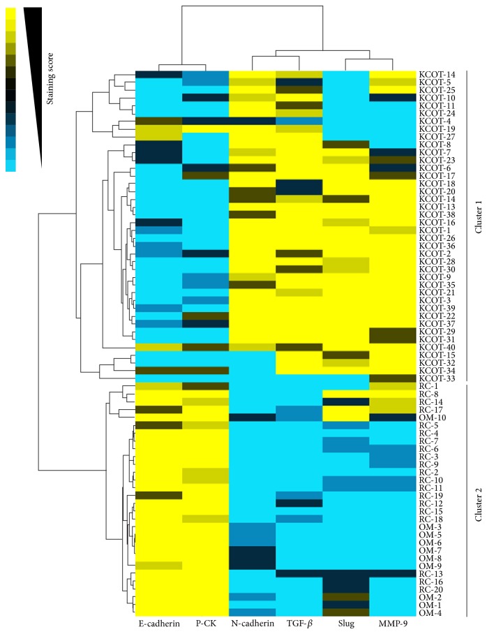
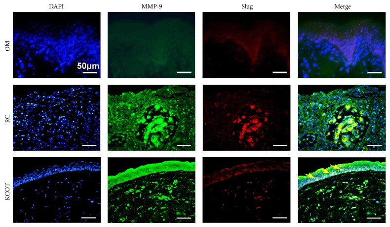
The aim of this study is to clarify whether epithelial-mesenchymal transition (EMT) is involved in the pathogenesis and development of keratocystic odontogenic tumor (KCOT). The expression levels of EMT-related proteins and genes in normal oral mucosa (OM), radicular cyst (RC), and KCOT were determined and compared by real-time quantitative PCR and immunohistochemistry. Our data showed that the expression of epithelial markers E-cadherin and Pan-cytokeratin was significantly downregulated in KCOT with upregulation of mesenchymal markers N-cadherin compared to OM and RC. Importantly, TGF-β, a potent EMT inducer, and Slug, a master transcription factor, were also found highly expressed in KCOT. In addition, the results from Spearman rank correlation test and clustering analysis revealed the close relationship between Slug and MMP-9, which was further evidenced by double-labeling immunofluorescence that revealed a synchronous distribution for Slug with MMP-9 in KCOT samples. All the data suggested EMT might be involved in the locally aggressive behavior of KCOT.


Figures

Figure 1
Figure 1
The mRNA expression levels of E-cadherin (a), N-cadherin (b), TGF-β (c), and Slug (d) in KCOT, RC, and OM tissue samples were detected by real-time qPCR analysis. Data are expressed as means accompanied by SEM. * P < 0.05 and *** P < 0.001.
Figure 2
Figure 2
(a) The immunohistochemical staining of E-cadherin, P-CK, N-cadherin, TGF-β, and Slug in KCOT, RC, and OM tissue samples. (b) Immunostaining scores for the tested EMT-related markers in KCOT, RC, and OM samples. Data are presented as means accompanied by SEM. *** P < 0.001.
Figure 3
Figure 3
Spearman rank test analyses for immunostaining of some EMT-related markers in KCOT samples. (a) The expression level of E-cadherin indicated a significantly positive correlation with P-CK. (b) The expression level of Slug showed a significantly positive correlation with TGF-β. (c) The expression level of Slug displayed a significantly negative correlation with E-cadherin.
Figure 4
Figure 4
Expression levels of MMP-9 in OM, RC, and KCOT samples, respectively, and correlation analyses for EMT-related markers and MMP-9. (a) The immunohistochemical staining of MMP-9 in KCOT, RC, and OM samples. (b) Immunostaining scores for MMP-9 in KCOT, RC, and OM samples. (c) The expression level of MMP-9 showed a significantly inverse correlation with E-cadherin. (d) The expression level of MMP-9 showed a significantly positive correlation with Slug. Data are presented as means accompanied by SEM. *** P < 0.001.
Figure 5
Figure 5
Clustering analysis for tested markers in 40 KCOT, 20 RC, and 10 OM samples. The relationship between indicated markers (top) was visually reflected by the length and subdivision of the branches in the heat-map. In addition, all of the KCOT cases (Cluster 1) and all of the RC and OM cases (Cluster 2) clustered together, revealing the significant difference.
Figure 6
Figure 6
Double-labeling immunofluorescence staining for Slug and MMP-9 in OM, RC, and KCOT tissue samples. The results showed a synchronous expression for Slug and MMP-9 in KCOT as the yellow arrows indicated.

References

    1. Thompson L. D. R. World Health Organization classification of tumours: pathology and genetics of head and neck tumours. Ear, Nose and Throat Journal. 2006;85(2):p. 74. - PubMed
    1. Sleeman J. P., Thiery J. P. SnapShot: the epithelial-mesenchymal transition. Cell. 2011;145(1):162–162.e1. doi: 10.1016/j.cell.2011.03.029. - DOI - PubMed
    1. Friedl P., Alexander S. Cancer invasion and the microenvironment: plasticity and reciprocity. Cell. 2011;147(5):992–1009. doi: 10.1016/j.cell.2011.11.016. - DOI - PubMed
    1. Jia J., Zhang W., Liu J.-Y., et al. Epithelial mesenchymal transition is required for acquisition of anoikis resistance and metastatic potential in adenoid cystic carcinoma. PLoS ONE. 2012;7(12) doi: 10.1371/journal.pone.0051549.e51549 - DOI - PMC - PubMed
    1. Tsuji T., Ibaragi S., Hu G.-F. Epithelial-mesenchymal transition and cell cooperativity in metastasis. Cancer Research. 2009;69(18):7135–7139. doi: 10.1158/0008-5472.can-09-1618. - DOI - PMC - PubMed

Publication types

MeSH terms

Substances

LinkOut - more resources