. 2015 Apr 16;10(4):e0123713.
doi: 10.1371/journal.pone.0123713. eCollection 2015.

Positively-charged semi-tunnel is a structural and surface characteristic of polyphosphate-binding proteins: an in-silico study

Affiliations

Positively-charged semi-tunnel is a structural and surface characteristic of polyphosphate-binding proteins: an in-silico study

Zheng Zachory Wei et al. PLoS One. .

Abstract

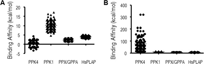
Phosphate is essential for all major life processes, especially energy metabolism and signal transduction. A linear phosphate polymer, polyphosphate (polyP), linked by high-energy phosphoanhydride bonds, can interact with various proteins, playing important roles as an energy source and regulatory factor. However, polyP-binding structures are largely unknown. Here we proposed a putative polyP binding site, a positively-charged semi-tunnel (PCST), identified by surface electrostatics analyses in polyP kinases (PPKs) and many other polyP-related proteins. We found that the PCSTs in varied proteins were folded in different secondary structure compositions. Molecular docking calculations revealed a significant value for binding affinity to polyP in PCST-containing proteins. Utilizing the PCST identified in the β subunit of PPK3, we predicted the potential polyP-binding domain of PPK3. The discovery of this feature facilitates future searches for polyP-binding proteins and discovery of the mechanisms for polyP-binding activities. This should greatly enhance the understanding of the many physiological functions of protein-bound polyP and the involvement of polyP and polyP-binding proteins in various human diseases.


Conflict of interest statement

Competing Interests: The authors have declared that no competing interests exist.

Figures

Fig 1
Fig 1. Model structures of Positively-Charged Semi-Tunnel (PCST).
(A) PPK4 (PDB ID: 3G3Q); (B) PPK1 (PDB ID: 1XDP); (C) PPX/GPPA (PDB ID: 1T6C); (D) HsPLAP (PDB ID: 1EW2). Protein structure (left) and protein surface graphic (right). Colors on graphics represent surface charges of the 3D structure of the protein. Blue = positive; Red = negative. Yellow double end arrows indicate observed strip of PCST.
Fig 2
Fig 2. Binding affinities of polyP to polyP positive control proteins.
PPK4 (PDB ID: 3G3Q); PPK1 (PDB ID:1XDO);PPX/GPPA (PDB ID: 1T6C); HsPLAP (PDB ID: 1EW2). PolyP ligands included Ap5 (A) and Ap6A (B). Each circle, square, or triangle represented an estimated free energy of binding from the molecular docking calculations using Ap5 or Ap6A ligand and different polyP positive control proteins.
Fig 3
Fig 3. Predicted binding energies of polyP to PPK partial structural analogues with or without a Positively-Charged Semi-Tunnel.
Each circle or square represented an estimated free energy of binding from the molecular docking calculations using Ap5 ligand and different PPK partial structural analogues.
Fig 4
Fig 4. PPK3 subunits and sequences.
(A) Multiple alignment of PPK3 subunit and actin amino acid sequences. Black arrows and blue highlights indicate strands; boxes and purple highlights indicate helices; yellow highlights indicate turns; inverted triangles and green highlights indicate nucleotide-binding regions. (B) The overlap of the backbone structures of the PPK3 subunits. α(blue); β(purple); ξ(green).
Fig 5
Fig 5. Binding affinities of polyP to subunits of PPK3.
Each circle, square, or triangle represented an estimated free energy of binding from the molecular docking calculations using Ap5 ligand and different PPK3 subunit.
Fig 6
Fig 6. PPK3 3D structure of subunits.
(A) β subunit; (B) ξ subunit; (C) α subunit. Protein structure (left) and protein surface graphic (right). Colors on graphics represent surface charges of the 3D structure of the protein. Blue = positive; Red = negative. Yellow double end arrows indicate observed strip of PCST.

References

    1. Lieberman L. (1888) Uber das Nuclein der Hefe und Kunstliche Darstellung eines Nucleus Eiweissund Metaphosphatsaure. Berichte der deutschen chemischen Gesellschaft 21: 598.
    1. Kumble KD, Kornberg A. (1995) Inorganic polyphosphate in mammalian cells and tissues. J Biol Chem 270: 5818–5822. - PubMed
    1. Dedkova EN, Blatter LA. (2014) Role of beta-hydroxybutyrate, its polymer poly-beta-hydroxybutyrate and inorganic polyphosphate in mammalian health and disease. Front Physiol 5: 260 10.3389/fphys.2014.00260 - DOI - PMC - PubMed
    1. Omelon S, Georgiou J, Henneman ZJ, Wise LM, Sukhu B, Hunt T, et al. (2009) Control of vertebrate skeletal mineralization by polyphosphates. PLoS One 4: e5634 10.1371/journal.pone.0005634 - DOI - PMC - PubMed
    1. Ariganello MB, Omelon S, Variola F, Wazen RM, Moffatt P, Nanci A, et al. (2014) Osteogenic cell cultures cannot utilize exogenous sources of synthetic polyphosphate for mineralization. J Cell Biochem 115: 2089–2102. 10.1002/jcb.24886 - DOI - PubMed

Publication types

LinkOut - more resources