Review
. 2015 May;265(1):6-21.
doi: 10.1111/imr.12296.

Regulation of inflammasome activation

Affiliations
Review

Regulation of inflammasome activation

Si Ming Man et al. Immunol Rev. 2015 May.

Abstract

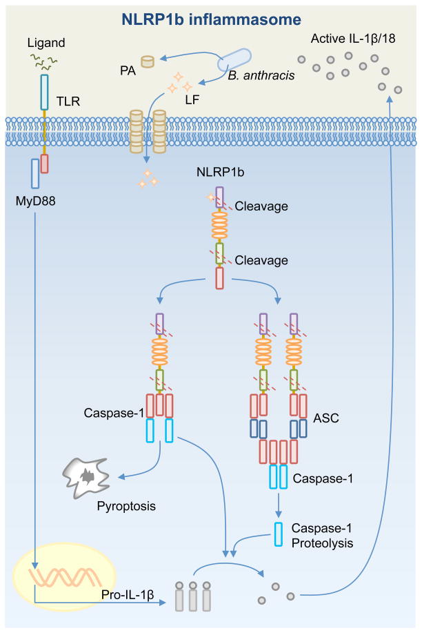
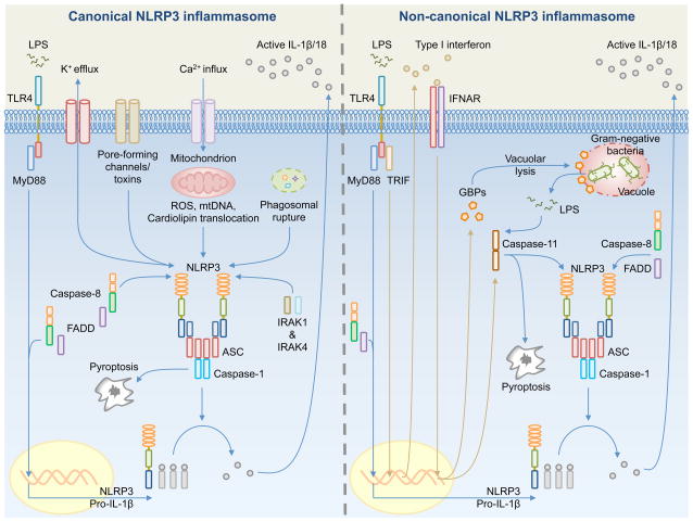
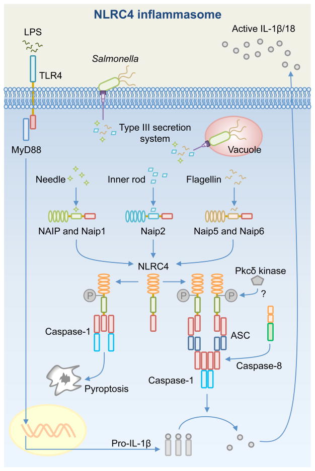
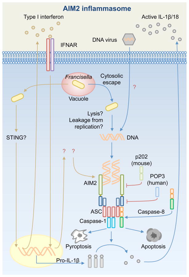
Inflammasome biology is one of the most exciting and rapidly growing areas in immunology. Over the past 10 years, inflammasomes have been recognized for their roles in the host defense against invading pathogens and in the development of cancer, auto-inflammatory, metabolic, and neurodegenerative diseases. Assembly of an inflammasome complex requires cytosolic sensing of pathogen-associated molecular patterns or danger-associated molecular patterns by a nucleotide-binding domain and leucine-rich repeat receptor (NLR) or absent in melanoma 2 (AIM2)-like receptors (ALR). NLRs and ALRs engage caspase-1, in most cases requiring the adapter protein apoptosis-associated speck-like protein containing a CARD (ASC), to catalyze proteolytic cleavage of pro-interleukin-1β (pro-IL-1β) and pro-IL-18 and drive pyroptosis. Recent studies indicate that caspase-8, caspase-11, IL-1R-associated kinases (IRAK), and receptor-interacting protein (RIP) kinases contribute to inflammasome functions. In addition, post-translational modifications, including ubiquitination, deubiquitination, phosphorylation, and degradation control almost every aspect of inflammasome activities. Genetic studies indicate that mutations in NLRP1, NLRP3, NLRC4, and AIM2 are linked with the development of auto-inflammatory diseases, enterocolitis, and cancer. Overall, these findings transform our understanding of the basic biology and clinical relevance of inflammasomes. In this review, we provide an overview of the latest development of inflammasome research and discuss how inflammasome activities govern health and disease.

Keywords: IL-1; NLR; caspase-1; caspase-11; caspase-8; inflammasome.


Conflict of interest statement

The authors have no conflicts of interest to declare.

Figures

Fig. 1
Fig. 1. An inflammasome complex can be visualized as a single distinct speck or focus in the cytoplasm of a cell
Primary mouse bone-marrow derived macrophages transfected with the dsDNA ligand poly(dA:dT) and stained for ASC (red), caspase-1 (green) and DNA (blue). Bar, 20 μM.
Fig. 2
Fig. 2. Activation of the NLRP1b inflammasome
Bacillus anthracis releases the anthrax lethal toxin, a bipartite toxin composed of a protective antigen (PA) and a lethal factor (LF). PA generates a pore on the host cell membrane, which is used by LF to enter the cell. Inflammasome responds to the presence of LF in the cytosol following LF-induced cleavage of NLRP1b at the N-terminal domain. Autoproteolytic cleavage at the FIIND domain of NLRP1b has also been observed. Cleavage of NLRP1b is sufficient to activate the inflammasome. In response to a high dose of LF, the CARD of NLRP1b binds the CARD of pro-caspase-1. This complex is sufficient to drive pro-IL-1β and pro-IL-18 processing and pyroptosis independently of ASC or caspases-1 self-proteolysis. In response to a low dose of LF, the CARD of NLRP1b recruits ASC to form a macromolecular cytoplasmic speck, where caspase-1 undergoes proteolysis and contributes to pro-IL-1β and pro-IL-18 processing.
Fig. 3
Fig. 3. Mechanisms of activation for the canonical and non-canonical NLRP3 inflammasomes
(Left). Canonical NLRP3 inflammasome activation requires priming by a Toll-like receptor (TLR) ligand (e.g. LPS) – mediated by MyD88, caspase-8, FADD, and NF-κB – to induce the expression of pro-IL-1β and NLRP3. Pro-IL-18 is expressed constitutively in the cell. A number of mechanisms have been proposed to activate the canonical NLRP3 inflammasome, including K+ efflux, pore-forming channels or toxins, Ca2+ influx, mitochondrial reactive oxygen species (ROS), mitochondrial DNA (mtDNA), translocation of cardiolipin from the inner mitochondrial membrane to the outer mitochondrial membrane, and phagosomal destabilization. NLRP3, ASC and caspase-1 assemble the inflammasome, which leads to proteolytic cleavage of pro-IL-1β and pro-IL-18 for release and the induction of pyroptosis. A priming-independent pathway for canonical NLRP3 inflammasome activation has been described. This pathway requires IRAK1 and IRK4. (Right). Non-canonical NLRP3 inflammasome activation is activated by Gram-negative bacteria. Extracellular LPS induces the expression of pro-IL-1β and NLRP3 via the TLR4-MyD88-dependent pathway and type I interferon via the TLR4-TRIF-dependent pathway. Type I interferon provides a feedback loop and activates type I interferon receptor (IFNAR) to induce caspase-11 expression. Cytosolic Gram-negative bacteria deliver LPS into the cytosol when they escape the vacuole. Vacuolar Gram-negative bacteria release their LPS into the cytosol through a mechanism that requires vacuolar rupture mediated by interferon-inducible guanylate-binding proteins (GBPs). Caspase-11 is proposed to activate following its binding to cytosolic LPS. Caspase-11 then drives pyroptosis and activation of the non-canonical NLRP3 inflammasome.
Fig. 4
Fig. 4. Activation of the NAIP-NLRC4 inflammasome
Toll-like receptors (e.g. TLR4) mediate the production of pro-IL-1β via MyD88 and NF-κB. Certain pathogenic bacteria use the Type III secretion system (T3SS) to deliver effector proteins to subvert host cell functions (e.g. Salmonella enterica serovar Typhimurium uses the Salmonella Pathogenicity Island-1 or SPI-1– Type III secretion system). In doing so, bacterial proteins such as the T3SS needle protein, inner rod protein, and flagellin are injected into the cytoplasm. These proteins are detected by NAIPs to activate the NLRC4 inflammasome, which results in pro-IL-1β and pro-IL-18 processing via an ASC-dependent mechanism. Caspase-1 and caspase-8 are recruited to the ASC inflammasome independently of each other during early infection. Caspase-8 is speculated to enhance delayed processing of pro-IL-1β and pro-IL-18 and induces delayed cell death. NLRC4 induces caspase-1-dependent pyroptosis via an ASC-independent mechanism. Phosphorylation of NLRC4 by the Pkcδ kinase is proposed to contribute to the activation of the NLRC4 inflammasome.
Fig. 5
Fig. 5. Activation of the AIM2 inflammasome
AIM2 is activated by cytosolic bacteria, such as Francisella tularensis, and DNA viruses, such as cytomegalovirus. During Francisella infection, type I interferon provides a feedback loop and activates type I interferon receptor (IFNAR). The downstream signaling of IFNAR is unknown. Francisella escapes the vacuole and replicates in the cytosol. It has been proposed that DNA release by means of bacteriolysis or bacterial replication in the cytosol activates AIM2. The mechanism leading to viral DNA recognition by AIM2 is less clear. The HIN-200 domain of AIM2 directly binds dsDNA and the pyrin domain recruits ASC. Caspase-1 and caspase-8 are recruited to the ASC inflammasome, where caspase-1 mediates pro-IL-1β and pro-IL-18 processing and pyroptosis and caspase-8 induces apoptosis. The pyrin-containing human protein POP3 and the HIN-domains-containing mouse p202 protein interact with AIM2 to inhibit inflammasome activation.

References

    1. Schroder K, Tschopp J. The inflammasomes. Cell. 2010;140:821–832. - PubMed
    1. Bertin J, et al. Human CARD4 protein is a novel CED-4/Apaf-1 cell death family member that activates NF-kappaB. The Journal of biological chemistry. 1999;274:12955–12958. - PubMed
    1. Inohara N, et al. Nod1, an Apaf-1-like activator of caspase-9 and nuclear factor-kappaB. The Journal of biological chemistry. 1999;274:14560–14567. - PubMed
    1. Harton JA, Linhoff MW, Zhang J, Ting JP. Cutting edge: CATERPILLER: a large family of mammalian genes containing CARD, pyrin, nucleotide-binding, and leucine-rich repeat domains. Journal of immunology. 2002;169:4088–4093. - PubMed
    1. Cridland JA, et al. The mammalian PYHIN gene family: phylogeny, evolution and expression. BMC evolutionary biology. 2012;12:140. - PMC - PubMed

Publication types