Review
. 2015 May;265(1):35-52.
doi: 10.1111/imr.12286.

Initiation and perpetuation of NLRP3 inflammasome activation and assembly

Affiliations
Review

Initiation and perpetuation of NLRP3 inflammasome activation and assembly

Eric I Elliott et al. Immunol Rev. 2015 May.

Abstract

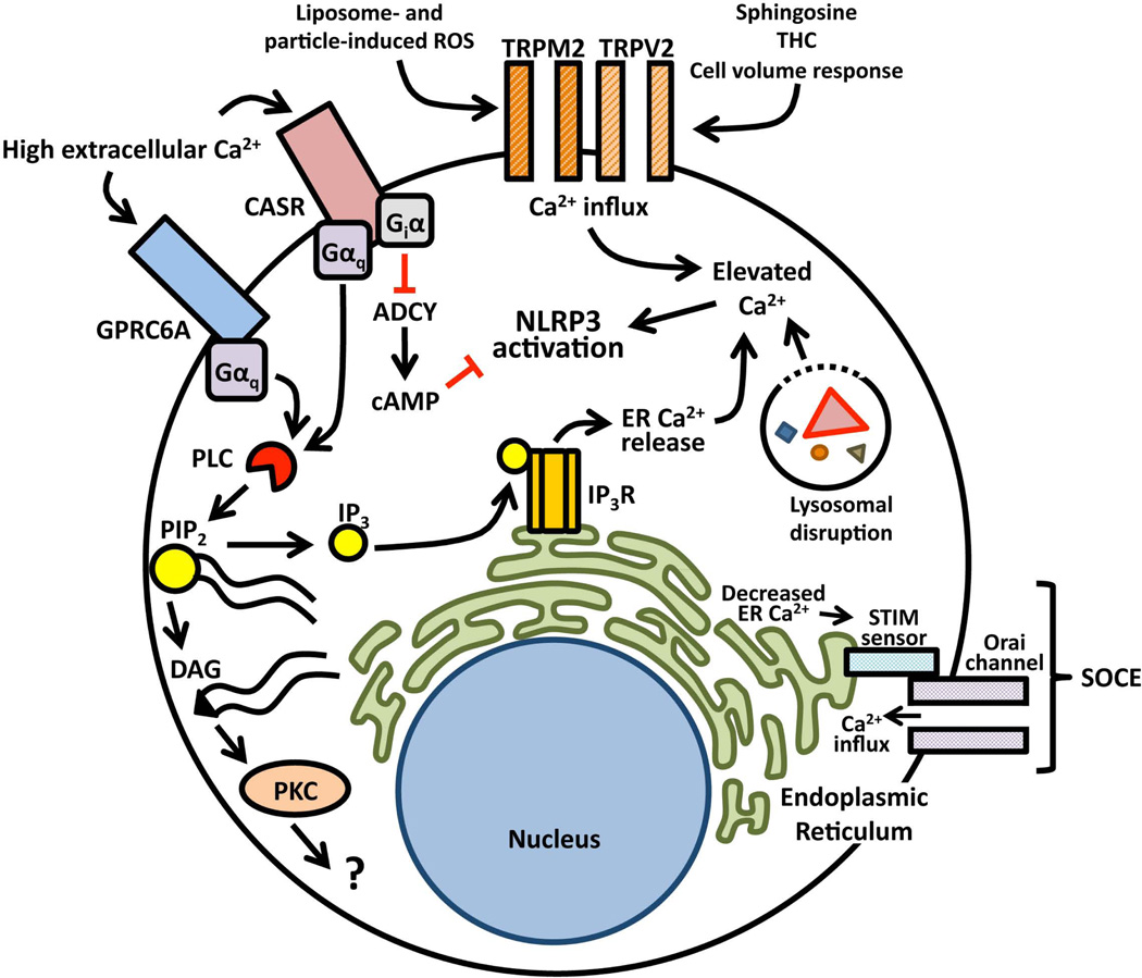
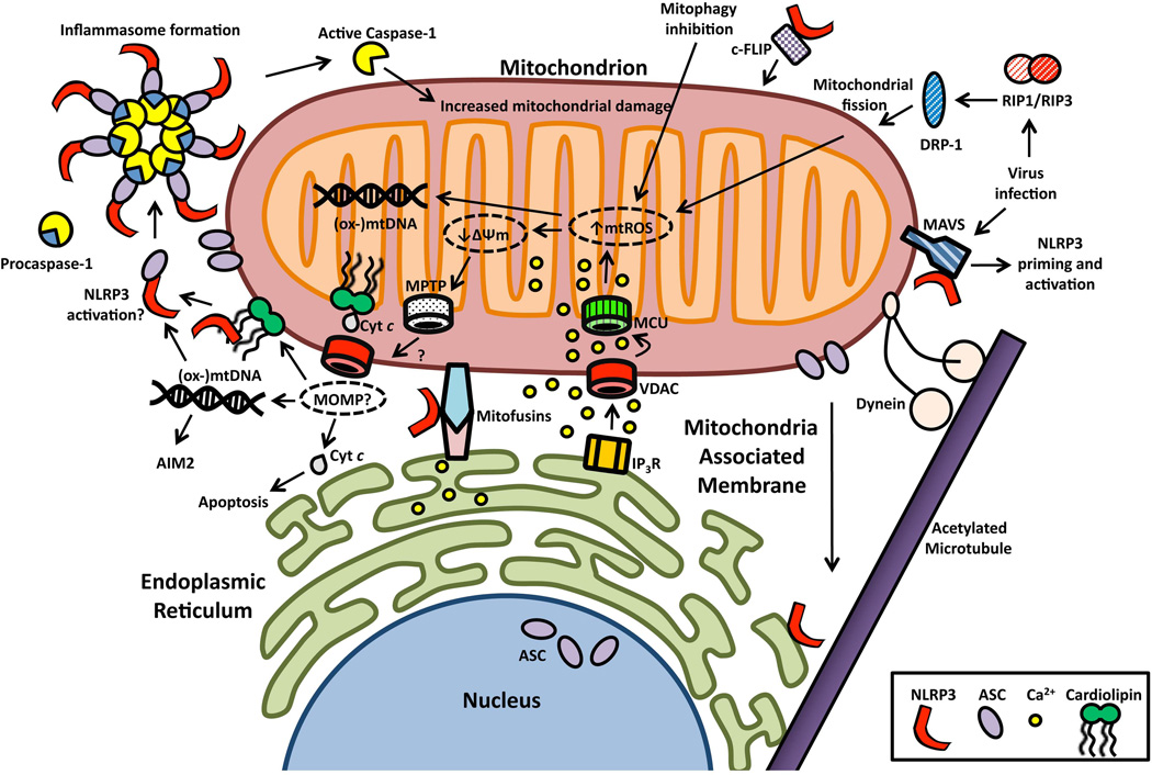
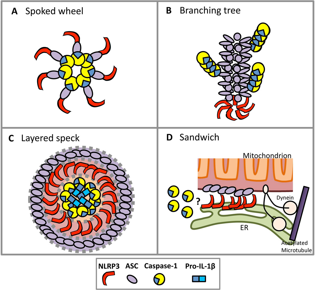
The NLRP3 (NOD-like receptor family, pyrin domain containing 3) inflammasome is a multiprotein complex that orchestrates innate immune responses to infection and cell stress through activation of caspase-1 and maturation of inflammatory cytokines pro-interleukin-1β (pro-IL-1β) and pro-IL-18. Activation of the inflammasome during infection can be protective, but unregulated NLRP3 inflammasome activation in response to non-pathogenic endogenous or exogenous stimuli can lead to unintended pathology. NLRP3 associates with mitochondria and mitochondrial molecules, and activation of the NLRP3 inflammasome in response to diverse stimuli requires cation flux, mitochondrial Ca(2+) uptake, and mitochondrial reactive oxygen species accumulation. It remains uncertain whether NLRP3 surveys mitochondrial integrity and senses mitochondrial damage, or whether mitochondria simply serve as a physical platform for inflammasome assembly. The structure of the active, caspase-1-processing NLRP3 inflammasome also requires further clarification, but recent studies describing the prion-like properties of ASC have advanced the understanding of how inflammasome assembly and caspase-1 activation occur while raising new questions regarding the propagation and resolution of NLRP3 inflammasome activation. Here, we review the mechanisms and pathways regulating NLRP3 inflammasome activation, discuss emerging concepts in NLRP3 complex organization, and expose the knowledge gaps hindering a comprehensive understanding of NLRP3 activation.

Keywords: NLRP3; caspase-1; inflammasome; mitochondria.


Conflict of interest statement

The authors have no conflicts of interest to declare.

Figures

Fig. 1
Fig. 1. Priming the NLRP3 inflammasome for activation
NLRP3 inflammasome priming is accomplished by NFκB-activating receptors including Toll-like receptors, interleukin-1 receptor, tumor necrosis factor receptor, and the cytosolic PRR NOD2. Nuclear translocation of NFκB leads to increased synthesis of NLRP3 and inflammasome-dependent cytokine pro-IL-1β. Priming also induces post-translational modifications to inflammasome components including NLRP3 deubiqutination and ASC ubiquitination and phosphorylation.
Fig. 2
Fig. 2. Activation of the NLRP3 inflammasome by particulate agonists
Extracellular particles and crystals are initially phagocytosed. Destabilization of the lysosome leads to release of lysosomal enzymes and calcium. It is unclear how release of these lysosomal molecules leads to NLRP3 activation, but it appears that lysosomal disruption induces cation flux through an unknown mechanism. It is also possible that phagolysosomal destabilization leads to activation of cell stress responsive kinases, which could affect upstream NLRP3 priming or inflammasome activation directly, but more work is necessary to clarify these mechanisms.
Fig. 3
Fig. 3. Regulation of NLRP3 activation by Ca2+ signaling
High extracellular Ca2+ from necrotic cells activates CASR and GPRC6A. Activation of these and other GPCRs leads to activation of PLC, which cleaves PIP2 into DAG and IP3. IP3 activates IP3R on ER membranes to trigger Ca2+ release. DAG activates PKC, but the role of this pathway in inflammasome activation is unclear. CASR also inhibits adenylate cyclase (ADCY), which may relieve the inhibition of NLRP3 by cAMP. Activation of NLRP3 requires SOCE, wherein STIM proteins sense ER Ca2+ depletion and trigger Ca2+ influx via Orai channels. NLRP3 inflammasome activation is also driven by Ca2+ uptake through TRPV2 and TRPM2 channels. Destabilized lysosomes could provide an additional source of Ca2+ for NLRP3 activation.
Fig. 4
Fig. 4. NLRP3 inflammasome activation is regulated by the mitochondrion
NLRP3 associates with mitochondria by interacting with MAVS, mitofusins, c-FLIP and cardiolipin. ER and mitochondrial membranes are tethered at the MAM via interactions between VDAC and IP3R as well as mitofusins. The IP3R releases ER Ca2+ stores while VDAC and MCU mediate mitochondrial Ca2+ uptake. Mitochondrial Ca2+ overload results in increased mtROS, which is required for NLRP3 activation. The role of mtROS in NLRP3 activation could be to increase mitochondrial damage or to create a DAMP by oxidizing mitochondrial molecules such as mtDNA. High mitochondrial Ca2+ and mtROS leads to loss of ΔΨm and MPTP, but it is unclear whether altered ΔΨm and MPTP are required for NLRP3 activation. MOMP permits release of cytochrome c from the inner-membrane space into the cytosol to interact with Apaf-1 for initiation of apoptosis, but the role of MOMP in NLRP3 activation is also uncertain. It is possible that MPTP or MOMP could regulate the release of a mitochondrial DAMP for NLRP3 such as (oxidized) mtDNA or cardiolipin. While the activating ligand of NLRP3 remains unknown, NLRP3 activation ultimately leads to formation of an inflammasome containing ASC and procaspase-1 and results in caspase-1 activation. Activation of caspase-1 in turn leads to increased mitochondrial damage. Inhibition of mitophagy prevents clearance of damaged mitochondria and increases mtROS and NLRP3 activation. Additionally, the NLRP3 response to viruses may involve priming and activation mediated by MAVS and RIP1/3-mediated activation of DRP-1 with subsequent mitochondrial fission and mtROS production. Finally, dynein-mediated mitochondrial transport along microtubules may regulate NLRP3 and ASC interactions.
Fig. 5
Fig. 5. Models of NLRP3 inflammasome complex assembly
(A) Traditionally, the inflammasome has been modeled after the apoptosome and thought to consist of peripheral NLRP3 spokes interacting indirectly with caspase-1 at the central hub through the adapter ASC. (B) The NLRP3 inflammasome resembles a tree where a heptameric NLRP3 root nucleates a fibrous ASC trunk, and caspase-1 recruited to the complex polymerizes laterally as branches. (C) The ASC speck is an inflammasome in which molecules of NLRP3 are contained within an ASC aggregate and caspase-1 and IL-1β are confined to the core of the complex. (D) The NLRP3 inflammasome is formed by the sandwich-like joining of ER membrane-associated NLRP3 with ASC on the outer mitochondrial membrane, which is regulated by dynein-mediated transport along microtubules.

References

    1. Ting JP, et al. The NLR gene family: a standard nomenclature. Immunity. 2008;28:285–287. - PMC - PubMed
    1. Khare S, et al. An NLRP7-containing inflammasome mediates recognition of microbial lipopeptides in human macrophages. Immunity. 2012;36:464–476. - PMC - PubMed
    1. Ataide MA, et al. Malaria-induced NLRP12/NLRP3-dependent caspase-1 activation mediates inflammation and hypersensitivity to bacterial superinfection. PLoS pathogens. 2014;10:e1003885. - PMC - PubMed
    1. Vladimer GI, et al. The NLRP12 inflammasome recognizes Yersinia pestis. Immunity. 2012;37:96–107. - PMC - PubMed
    1. Kumar H, et al. NLRC5 deficiency does not influence cytokine induction by virus and bacteria infections. Journal of immunology. 2011;186:994–1000. - PubMed

Publication types