. 2015 Mar 7:12:45.
doi: 10.1186/s12974-015-0237-4.

Transiently lowering tumor necrosis factor-α synthesis ameliorates neuronal cell loss and cognitive impairments induced by minimal traumatic brain injury in mice

Affiliations

Transiently lowering tumor necrosis factor-α synthesis ameliorates neuronal cell loss and cognitive impairments induced by minimal traumatic brain injury in mice

Renana Baratz et al. J Neuroinflammation. .

Abstract

Background: The treatment of traumatic brain injury (TBI) represents an unmet medical need, as no effective pharmacological treatment currently exists. The development of such a treatment requires a fundamental understanding of the pathophysiological mechanisms that underpin the sequelae resulting from TBI, particularly the ensuing neuronal cell death and cognitive impairments. Tumor necrosis factor-alpha (TNF-α) is a cytokine that is a master regulator of systemic and neuroinflammatory processes. TNF-α levels are reported to become rapidly elevated post TBI and, potentially, can lead to secondary neuronal damage.

Methods: To elucidate the role of TNF-α in TBI, particularly as a drug target, the present study evaluated (i) time-dependent TNF-α levels and (ii) markers of apoptosis and gliosis within the brain and related these to behavioral measures of 'well being' and cognition in a mouse closed head 50 g weight drop mild TBI (mTBI) model in the presence and absence of post-treatment with an experimental TNF-α synthesis inhibitor, 3,6'-dithiothalidomide.

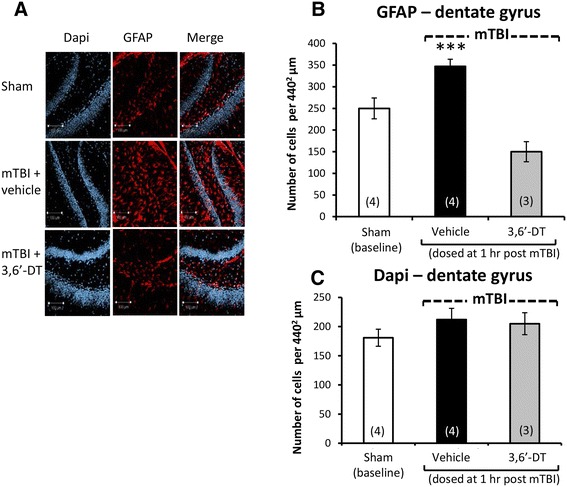
Results: mTBI elevated brain TNF-α levels, which peaked at 12 h post injury and returned to baseline by 18 h. This was accompanied by a neuronal loss and an increase in astrocyte number (evaluated by neuronal nuclei (NeuN) and glial fibrillary acidic protein (GFAP) immunostaining), as well as an elevation in the apoptotic death marker BH3-interacting domain death agonist (BID) at 72 h. Selective impairments in measures of cognition, evaluated by novel object recognition and passive avoidance paradigms - without changes in well being, were evident at 7 days after injury. A single systemic treatment with the TNF-α synthesis inhibitor 3,6'-dithiothalidomide 1 h post injury prevented the mTBI-induced TNF-α elevation and fully ameliorated the neuronal loss (NeuN), elevations in astrocyte number (GFAP) and BID, and cognitive impairments. Cognitive impairments evident at 7 days after injury were prevented by treatment as late as 12 h post mTBI but were not reversed when treatment was delayed until 18 h.

Conclusions: These results implicate that TNF-α in mTBI induced secondary brain damage and indicate that pharmacologically limiting the generation of TNF-α post mTBI may mitigate such damage, defining a time-dependent window of up to 12 h to achieve this reversal.


Figures

Figure 1
Figure 1
mTBI induces a time-dependent rise in brain TNF-α levels. Right (ipsilateral to mTBI) cerebral cortex protein extracts were prepared from sham or mTBI mice at the indicated time points post injury. (A) Time-dependent brain levels of TNF-α at baseline (sham) and post injury. At 12 h post mTBI, TNF-α levels peaked (132.8 vs. 53.4 (sham) pg/ml, p < 0.0001). By 18 h post injury, TNF-α levels returned to baseline (50.5 pg/ml). (B) Treatment with 3,6′-dithiothalidomide (3,6-DT) at 1 h after mTBI prevented the TNF-α elevation evident at 12 h post mTBI (3,6′-DT + mTBI 67.1 pg/ml vs. mTBI 132.8 pg/ml, p < 0.0001). In both (A) and (B), **** was significantly different from all other groups (p < 0.0001).
Figure 2
Figure 2
mTBI induces impairments in performance in both a Y-maze and novel object recognition (NOR) preference index paradigms that are ameliorated by 3,6′-dithiothalidomide when administered up to 12 but not 18 h post injury. (A) Performance of mice was quantitatively assessed in a Y-maze and (B) in a NOR paradigm at 7 days following mTBI as a preference index that was calculated as (time associated with the novel − time with the old arm or object)/(time with the novel + time with the old arm or object). Values are mean ± SEM values; a one-way ANOVA indicates that mTBI animals had a deficit in spatial (Y-maze) and visual (NOR) memory performance compared with all the other groups (*p < 0.05) with the exception of animals dosed with 3,6′-dithiothalidomide at 18 h post injury. No differences were found between any of the other groups (control (sham) 1 and 12 h 3,6′-dithiothalidomide dosing), suggesting complete amelioration by 3,6′-dithiothalidomide when administered within 12 h of injury.
Figure 3
Figure 3
Neuronal loss and apoptosis is induced by mTBI in cerebral cortex ipsilateral to injury and mitigated by 3,6′-dithiothalidomide. At 72 h post injury, cerebral cortex ipsilateral to mTBI was assessed for cellular changes. (A) and (B) A decline in neuronal number indicative of neuronal loss (NeuN - green) was evident post mTBI (p < 0.01). Treatment with 3,6′-dithiothalidomide at 1 h post-injury prevented such a change. (A) and (C) An elevation in BID (a marker for apoptosis - red) was evident within mTBI brains (p < 0.001). No changes in apoptotic cell death were found in animals that were treated with 3,6′-dithiothalidomide (as compared to sham animals). Within (A) (representative sections within the cerebral cortex), the bar is equal to 20 μm in length.
Figure 4
Figure 4
Neuronal loss and apoptosis is induced by mTBI in the dentate gyrus ipsilateral to injury and mitigated by 3,6′-dithiothalidomide. At 72 h post injury, the dentate gyrus of the hippocampus ipsilateral to mTBI was evaluated for cellular changes. (A) and (B) Neuronal loss (NeuN - green) was found post mTBI (p < 0.05). Treatment with the 3,6′-dithiothalidomide at 1 h post-injury prevented this loss. (A) and (C) An increase in BID (a marker for apoptosis in red) was evident in the mTBI brains (p < 0.01). No change in apoptotic cell death was apparent in animals treated with 3,6′-dithiothalidomide (as compared to sham animals). Within (A) (representative sections within the dentate gyrus), the bar is equal to 100 μm in length.
Figure 5
Figure 5
mTBI induces an elevation in astrocyte number in ipsilateral cerebral cortex that is inhibited by 3,6′-dithiothalidomide. At 72 h post injury, cerebral cortex ipsilateral to mTBI was assessed for cellular changes. (A) and (B) Astrocyte number (GFAP - red) was increased post mTBI (p < 0.001). Treatment with 3,6′-dithiothalidomide at 1 h post-injury prevented this. (A) and (C) No difference in total number of cells was evident between groups, as revealed from DAPI (blue) staining. Within (A) (representative sections within the cerebral cortex), the bar is equal to 100 μm in length.
Figure 6
Figure 6
mTBI induces an elevation in astrocyte number in ipsilateral dentate gyrus that is inhibited by 3,6′-dithiothalidomide. At 72 h post injury, dentate gyrus ipsilateral injury was assessed for cellular changes. (A) and (B) Astrocyte number (GFAP - red) was elevated post mTBI (p < 0.001). Treatment with 3,6′-dithiothalidomide at 1 h post-injury inhibited this. (A) and (C) No difference in total number of cells was apparent between groups, as evaluated by DAPI (blue) staining. Within (A) (representative sections within the dentate gyrus), the bar is equal to 100 μm in length.

References

    1. Hyder AA, Wunderlich CA, Puvanachandra P, Gururaj G, Kobusingye OC. The impact of traumatic brain injuries: a global perspective. NeuroRehabilitation. 2007;22:341–53. - PubMed
    1. Setnik L, Bazarian JJ. The characteristics of patients who do not seek medical treatment for traumatic brain injury. Brain Inj. 2007;21(1):1–9. doi: 10.1080/02699050601111419. - DOI - PubMed
    1. Faul M, Xu L, Wald M, Coronado VG. Traumatic Brain Injury in the United States: Emergency Department Visits, Hospitalizations and Deaths 2002–2006. Atlanta (GA): Centers for Disease Control and Prevention, National Center for Injury Prevention and Control; 2010.
    1. McGarry LJ, Thompson D, Millham FH, Cowell L, Snyder PJ, Lenderking WR, et al. Outcomes and costs of acute treatment of traumatic brain injury. J Trauma. 2002;53(6):1152–9. doi: 10.1097/00005373-200212000-00020. - DOI - PubMed
    1. McAllister TW. Neurobehavioral sequelae of traumatic brain injury: evaluation and management. World Psychiatry. 2008;7(1):3–10. - PMC - PubMed

Publication types

MeSH terms