. 2015 Mar 6:12:40.
doi: 10.1186/s12985-015-0266-8.

BRCA1 functions as a novel transcriptional cofactor in HIV-1 infection

Affiliations

BRCA1 functions as a novel transcriptional cofactor in HIV-1 infection

Irene Guendel et al. Virol J. .

Abstract

Background: Viruses have naturally evolved elegant strategies to manipulate the host's cellular machinery, including ways to hijack cellular DNA repair proteins to aid in their own replication. Retroviruses induce DNA damage through integration of their genome into host DNA. DNA damage signaling proteins including ATR, ATM and BRCA1 contribute to multiple steps in the HIV-1 life cycle, including integration and Vpr-induced G2/M arrest. However, there have been no studies to date regarding the role of BRCA1 in HIV-1 transcription.

Methods: Here we performed various transcriptional analyses to assess the role of BRCA1 in HIV-1 transcription by overexpression, selective depletion, and treatment with small molecule inhibitors. We examined association of Tat and BRCA1 through in vitro binding assays, as well as BRCA1-LTR association by chromatin immunoprecipitation.

Results: BRCA1 was found to be important for viral transcription as cells that lack BRCA1 displayed severely reduced HIV-1 Tat-dependent transcription, and gain or loss-of-function studies resulted in enhanced or decreased transcription. Moreover, Tat was detected in complex with BRCA1 aa504-802. Small molecule inhibition of BRCA1 phosphorylation effector kinases, ATR and ATM, decreased Tat-dependent transcription, whereas a Chk2 inhibitor showed no effect. Furthermore, BRCA1 was found at the viral promoter and treatment with curcumin and ATM inhibitors decreased BRCA1 LTR occupancy. Importantly, these findings were validated in a highly relevant model of HIV infection and are indicative of BRCA1 phosphorylation affecting Tat-dependent transcription.

Conclusions: BRCA1 presence at the HIV-1 promoter highlights a novel function of the multifaceted protein in HIV-1 infection. The BRCA1 pathway or enzymes that phosphorylate BRCA1 could potentially be used as complementary host-based treatment for combined antiretroviral therapy, as there are multiple potent ATM inhibitors in development as chemotherapeutics.


Figures
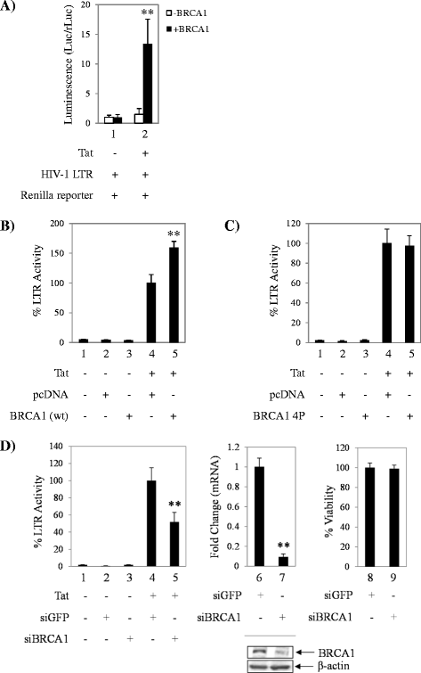
Figure 1
Figure 1
HIV-1 Tat-dependent transcription is more efficient in cells containing BRCA1. A. UWB1.289 BRCA1 null and UWB1.289 + BRCA1 cells and were co-transfected with pcDNA or pcTat, LTR-Luc, and CMV-Luc (Renilla) plasmid DNA. Dual-Glo luciferase assay was performed 48 hours later as described by the manufacturer. Raw data was normalized to Renilla luciferase expression in both cell lines, fold changes were calculated against pcDNA (−Tat) condition for each cell line. B. TZM-bl cells were co-transfected with pcDNA or pcTat, and BRCA1 (wild-type) plasmid DNA. Bright-Glo luciferase assay was performed 48 hours pos-transfection as described by the manufacturer. Cells containing pcTat and pcDNA were used as baseline value for Tat-dependent LTR activation. C. TZM-bl cells were co-transfected with pcDNA or pcTat, and BRCA1 4P (mutant) plasmid DNA (S1387A, S1423A, S1457A, and S1524A). Bright-Glo luciferase assay was performed 48 hours post-transfection. Cells containing pcTat and pcDNA were used as baseline value for Tat-dependent LTR activation. D. TZM-bl cells were co-transfected with pcTat or siRNA against GFP (control) and BRCA1. Bright-Glo luciferase assay was performed 48 hours post-transfection (left panel). Cells containing pcTat and siGFP were used as baseline value for Tat-dependent LTR activation. BRCA1 depletion was confirmed by qRT-PCR and western blot (middle panel and inset). Fold changes against siGFP were calculated relative to Actin using the ΔΔCt method. CellTiter-Glo cell viability assay (right panel) was performed as described by the manufacturer and viability normalized to cells transfected with siGFP. Transfection assays were performed in triplicate and data plotted represents averaged data of two independent experiments. Error bars show the standard error of two averaged independent measurements. Viability assays were performed in triplicate. Western blots were performed for two independent experiments. Double asterisk indicates statistically significant difference p ≤ 0.01.
Figure 2
Figure 2
BRCA1 associates with Tat. A. Schematic of five GST-BRCA1 fragments spanning the whole length protein. B. TZM-bl cells were transfected with Flag-Tat plasmid DNA for 48 hours. One milligram of whole cell protein extract was incubated with 1 μg GST-BRCA1 constructs overnight and processed as described in the Methods section. Samples were analyzed by western blot (top panel WB: anti-Flag, bottom panel WB: anti-BRCA1). C. TZM-bl cells were transfected with Flag-Tat plasmid DNA for 48 hours. One milligram of TZM-bl whole cell protein extract was immunoprecipitated with anti-BRCA1 and anti-IgG antibodies and analyzed by western blot (top panel WB: anti-BRCA1, middle panel WB: anti-Flag, bottom panel WB: anti-BRG1). Western blots are representative of two independent experiments.
Figure 3
Figure 3
BRCA1 status affects enhancement of Tat-dependent transcription during infection with pseudotyped particles. A. Schematic depicting the LTR-driven reporter plasmid pNL-RRE-SA-Luc. B. UWB1.289 BRCA1 null and UWB1.289 + BRCA1 cells and were co-transfected with pcTat and CMV-Luc (Renilla) plasmid DNA 24 hours prior to infection with LTR-driven reporter viral particles. Dual-Glo luciferase assay was performed 24 hours post-infection as described by the manufacturer. Raw data was normalized to Renilla luciferase expression in both cell lines, fold changes were calculated against + Tat mock infected cells (lane 1). Transfection and infection assays were performed in triplicate and data plotted represents averaged data of two independent experiments. Single asterisk indicates p < 0.05. C. UWB1.289 BRCA1 null and UWB1.289 + BRCA1 cells were infected with VSVG-pseudotyped NL4-3. Cells were collected 48 hours post-infection, stained with propidium iodine, and analyzed by flow cytometry. Results are representative of three biological replicates. D. Cells were infected as described above and collected at 48 hours post-infection for ChIP analysis. Antibodies used for ChIP were anti-RNA polymerase II (RNAP II, 10 μg), anti-Sp1 (10 μg), and anti-V5 (10 μg). Quantitative PCR was performed using SYBR Green PCR Master Mix to analyze immunoprecipitated material.
Figure 4
Figure 4
Curcumin decreases BRCA1 occupancy at the HIV-1 LTR. A. TZM-bl cells were transfected with pcTat and treated the next day with vehicle (DMSO) or a titration of curcumin (0.5, 1, 10, and 20 μM). Samples were analyzed by western blot. Inset depicts a dose–response curve of BRCA1 protein abundance versus curcumin treatment based on the average densitometry counts with error bars representing standard error of three independent measurements. Western blot is representative of three independent experiments. Densitometry counts were taken from three independent treatments to acquire a dose–response curve of BRCA1 expression inhibition (inset plot) B. TZM-bl cells were transfected with pcTat and treated the next day with DMSO or 20 μM curcumin. Bright-Glo luciferase assays and CellTiter-Glo cell viability assays were performed 24 hours post-treatment as described by the manufacturer. Data was normalized to cells containing Tat and treated with DMSO as baseline for Tat-dependent LTR activation. Transfection and treatment assays were performed in triplicate and data plotted represents averaged data of two independent experiments. Error bars show the standard error of two averaged independent measurements. Viability assays were performed in triplicate. C. TZM-bl cells were transfected with pcTat and treated the next day with DMSO or curcumin (20 μM) for 24 hours prior to being collected for ChIP analysis. Antibodies used for ChIP were anti-BRCA1 (10 μg), anti-IgG (10 μg), and anti-RNA polymerase II (RNAP II, 10 μg). Quantitative PCR was performed using SYBR Green PCR Master Mix to analyze immunoprecipitated material. Single asterisk indicates p < 0.05 and double asterisk indicates statistically significant difference p ≤ 0.01.
Figure 5
Figure 5
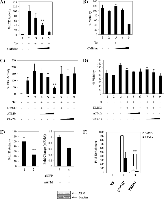
Inhibition of upstream BRCA1 phosphorylation effectors ATR/ATM, decreases Tat-dependent transcription. A. TZM-bl cells were transfected with pcTat and treated the next day with vehicle (water) and a titration of caffeine (500 μM, 2 mM and 5 mM). Bright-Glo luciferase assays were performed 48 hours post-treatment. Data was normalized to cells containing Tat and treated with DMSO as baseline for Tat-dependent LTR activation. B. CellTiter-Glo cell viability assays were performed 48 hours post-treatment. Data was normalized as in panel A. C. TZM-bl cells were transfected with pcTat and treated the next day with vehicle (DMSO) and a titration of ATM or Chk2 inhibitors (ATMin and Chk2in) at 0.1 μM, 1 μM and 10 μM. Bright-Glo luciferase assays were performed 48 hours post-treatment. Data was normalized as in panel A. D. CellTiter-Glo cell viability assays were performed 48 hours post-treatment. Data was normalized as in panel A. E. TZM-bl cells were co-transfected with pcTat or siRNA against GFP (control) and ATM. Bright-Glo luciferase assays were performed 48 hours post-treatment. Cells containing pcTat and siGFP were used as baseline value for Tat-dependent LTR activation. ATM depletion was confirmed by qRT-PCR and western blot (right panel and inset). Fold changes against siGFP were calculated relative to Actin using the ΔΔCt method. F. TZM-bl cells were transfected with pcTat and treated the next day with DMSO or ATM inhibitor (10 μM) for 48 hours prior to being collected for ChIP analysis. Antibodies used for ChIP were anti-BRCA1 (10 μg), anti-V5 (10 μg), and anti- pS10-H3 (10 μg). Transfection and treatment assays were performed in triplicate and data represents averaged data of two independent experiments. Viability assays were performed in triplicate. Error bars show the standard error of two averaged independent measurements. Double asterisk indicates statistically significant difference p ≤ 0.01.
Figure 6
Figure 6
BRCA1 is present at the HIV-1 LTR in HIV infected T-cells. A. CEM cells were infected with NL4-3 virus (p24 = 5000 pg/ml) for 4 hours and collected 72 hours post-infection for ChIP analysis. Antibodies used for ChIP were anti-BRCA1 (10 μg), anti-V5 (10 μg), and anti-histone H3-phosphorylated at S10 (pS10-H3, 5 μg). Quantitative PCR was performed using SYBR Green PCR Master Mix to analyze immunoprecipitated material. B. CEM cells were pre-treated with DMSO or 10 μM ATM inhibitor (ATMin) for 2 hours. Cells were then infected with NL4-3 virus (p24 = 5000 pg/ml) for 4 hours, followed by post-treating the cells with DMSO or ATMin. Cells were collected 72 hours post-infection for ChIP analysis. Antibodies used for ChIP were anti-BRCA1 (10 μg), p-BRCA1 S1423 (10 μg), and anti-V5 (10 μg). Quantitative PCR was performed using SYBR Green PCR Master Mix to analyze immunoprecipitated material. Double asterisk indicates statistically significant difference p ≤ 0.01.

Similar articles

Cited by

References

    1. Gatignol A. Transcription of HIV: Tat and cellular chromatin. Adv Pharmacol. 2007;55:137–59. doi: 10.1016/S1054-3589(07)55004-0. - DOI - PubMed
    1. Peng J, Zhu Y, Milton JT, Price DH. Identification of multiple cyclin subunits of human P-TEFb. Genes Dev. 1998;12:755–62. doi: 10.1101/gad.12.5.755. - DOI - PMC - PubMed
    1. Fujinaga K, Cujec TP, Peng J, Garriga J, Price DH, Grana X, et al. The ability of positive transcription elongation factor B to transactivate human immunodeficiency virus transcription depends on a functional kinase domain, cyclin T1, and Tat. J Virol. 1998;72:7154–9. - PMC - PubMed
    1. Herrmann CH, Rice AP. Lentivirus Tat proteins specifically associate with a cellular protein kinase, TAK, that hyperphosphorylates the carboxyl-terminal domain of the large subunit of RNA polymerase II: candidate for a Tat cofactor. J Virol. 1995;69:1612–20. - PMC - PubMed
    1. Laspia MF, Rice AP, Mathews MB. HIV-1 Tat protein increases transcriptional initiation and stabilizes elongation. Cell. 1989;59:283–92. doi: 10.1016/0092-8674(89)90290-0. - DOI - PubMed

Publication types

MeSH terms

Substances