. 2015 May;30(6):834-42.
doi: 10.1002/mds.26231. Epub 2015 Apr 16.

Oscillatory head movements in cervical dystonia: Dystonia, tremor, or both?

Affiliations

Oscillatory head movements in cervical dystonia: Dystonia, tremor, or both?

Aasef G Shaikh et al. Mov Disord. 2015 May.

Abstract

Cervical dystonia is characterized by abnormal posturing of the head, often combined with tremor-like oscillatory head movements. The nature and source of these oscillatory head movements is controversial, so they were quantified to delineate their characteristics and develop a hypothetical model for their genesis. A magnetic search coil system was used to measure head movements in 14 subjects with cervical dystonia. Two distinct types of oscillatory head movements were detected for most subjects, even when they were not clinically evident. One type had a relatively large amplitude and jerky irregular pattern, and the other had smaller amplitude with a more regular and sinusoidal pattern. The kinematic properties of these two types of oscillatory head movements were distinct, although both were often combined in the same subject. Both had features suggestive of a defect in a central neural integrator. The combination of different types of oscillatory head movements in cervical dystonia helps to clarify some of the current debates regarding whether they should be considered as manifestations of dystonia or tremor and provides novel insights into their potential pathogenesis.

Keywords: basal ganglia; cerebellum; midbrain; neural integrator; torticollis.


Conflict of interest statement

Relevant conflicts of interest/financial disclosures: Nothing to report. Full financial disclosures and author roles may be found in the online version of this article.

Figures

FIG. 1
FIG. 1
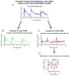
An example of oscillatory head movements from a subject with cervical dystonia attempting to hold the head steady. Head positions are plotted on the y-axis, and time is plotted on the x-axis. (A) The raw trace of head position shows two types, with small regular oscillatory head movements superimposed on larger irregular oscillatory head movements. (B) Sinusoidal oscillatory head movements was separated from jerky oscillatory head movements by using low-pass filtering. Green trace illustrates low-pass filtered waveform that characterizes jerky oscillatory head movements. (C) Epochs of high-pass filtered small oscillatory head movements separated from the same epoch showed in this panel. (D) Detrended signal was aligned to zero by de-trending and offsetting by its mean value. The x-coordinate of the intercept between composite vector (red trace) moving from high to low and zero-line (black dashed line) is acquired (zero-crossing, green symbols in the figure). The difference between such zero-crossing assessed cycle width (or period) and the inverse of the period is frequency of the given cycle. The amplitude of the cycle was computed by measuring the difference between peak to trough (ie, difference between blue symbols in the figure). [Color figure can be viewed in the online issue, which is available at wileyonlinelibrary.com]
FIG. 2
FIG. 2
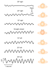
An example from a subject with cervical dystonia aiming the head in different positions to show the effect of different head positions on the amplitude of the small sinusoidal oscillatory head movements. Traces depict head position versus corresponding time. Positive deflections are rightward movements, whereas negative deflections are leftward movements. Caricatures next to trace depict the degree of head turn on the trunk. [Color figure can be viewed in the online issue, which is available at wileyonlinelibrary.com]
FIG. 3
FIG. 3
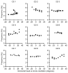
Position-dependent amplitudes of sinusoidal oscillatory head movements for all nine cervical dystonia subjects. The mean amplitudes of oscillatory head movements during 20-s epochs are plotted on the y-axis, whereas head orientations are plotted on the x-axis. Each data point depicts one observation over 20 s, and each panel illustrates one subject. The amplitude of oscillations changes with the head-on-trunk orientation. The intercept of the linear fit through the data depicts the “null” position for a given patient. The dashed vertical line depicts the location of the head on trunk at the time of maximal attenuation of dystonic tremor.
FIG. 4
FIG. 4
Example of head-on-trunk orientation dependence of the frequency of sinusoidal oscillatory head movements. Mean frequency 20-s epochs of eccentric head-on-trunk orientation is plotted on the y-axis, whereas corresponding head orientation is plotted on the x-axis. Each data point depicts one observation over 20 s, and each panel illustrates one subject. The frequency of oscillations remains insensitive to the head-on-trunk orientation.
FIG. 5
FIG. 5
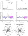
Influence of rapid voluntary horizontal head movement on oscillatory head movements in subjects with cervical dystonia. Panels A and C depict horizontal and vertical head positions, respectively, whereas panels B and D depict corresponding head velocities. Head positions and velocities are plotted on the y-axis, and the x-axis depicts corresponding time in seconds. Panels A and B depict the effects of voluntary horizontal head movement on the phase of oscillations. The phase was computed by determining the shift in the phase of sinusoidal fit in head velocity trace before voluntary head movement (blue dashed line) and after the voluntary head movement (magenta line). The same analysis is done for the effect of horizontal head movement on vertical head oscillations. Thus, phase shift after rapid horizontal head movement is measured for (on-axis) horizontal and (cross-axis) vertical head oscillations. Polar histograms show that in all instances there was a phase shift. Bins of the phase shift are plotted on the perimeter of the circle, and the bars at the spokes of the circle depict the number of instances that fall in a range of phase shift depicted by a given bin. D represents the summary of the phase shift of horizontal oscillations, whereas E is a phase shift of vertical oscillations. [Color figure can be viewed in the online issue, which is available at wileyonlinelibrary.com]

References

    1. Jinnah HA, Berardelli A, Comella C, et al. The focal dystonias: current views and challenges for future research. Mov Disord. 2013;28:926–943. - PMC - PubMed
    1. Chan J, Brin MF, Fahn S. Idiopathic cervical dystonia: clinical characteristics. Mov Disord. 1991;6:119–126. - PubMed
    1. Pal PK, Samii A, Schulzer M, Mak E, Tsui JK. Head tremor in cervical dystonia. Can J Neurol Sci. 2000;27:137–142. - PubMed
    1. Valls-Sole J, Tolosa ES, Nobbe F, et al. Neurophysiological investigations in patients with head tremor. Mov Disord. 1997;12:576–584. - PubMed
    1. Jankovic J, Leder S, Warner D, Schwartz K. Cervical dystonia: clinical findings and associated movement disorders. Neurology. 1991;41:1088–1091. - PubMed

Publication types