. 2015 Apr 16:8:155.
doi: 10.1186/s13104-015-1117-9.

Potential complications when developing gene deletion clones in Xylella fastidiosa

Affiliations

Potential complications when developing gene deletion clones in Xylella fastidiosa

Kameka L Johnson et al. BMC Res Notes. .

Abstract

Background: The Gram-negative xylem-limited bacterium, Xylella fastidiosa, is an important plant pathogen that infects a number of high value crops. The Temecula 1 strain infects grapevines and induces Pierce's disease, which causes symptoms such as scorching on leaves, cluster collapse, and eventual plant death. In order to understand the pathogenesis of X. fastidiosa, researchers routinely perform gene deletion studies and select mutants via antibiotic markers.

Methods: Site-directed pilJ mutant of X. fastidiosa were generated and selected on antibiotic media. Mutant cultures were assessed by PCR to determine if they were composed of purely transformant cells or included mixtures of non-transformants cells. Then pure pilJ mutant and wildtype cells were mixed in PD2 medium and following incubation and exposure to kanamycin were assessed by PCR for presence of mutant and wildtype populations.

Results: We have discovered that when creating clones of targeted mutants of X. fastidiosa Temecula 1 with selection on antibiotic plates, X. fastidiosa lacking the gene deletion often persist in association with targeted mutant cells. We believe this phenomenon is due to spontaneous antibiotic resistance and/or X. fastidiosa characteristically forming aggregates that can be comprised of transformed and non-transformed cells. A combined population was confirmed by PCR, which showed that targeted mutant clones were mixed with non-transformed cells. After repeated transfer and storage the non-transformed cells became the dominant clone present.

Conclusions: We have discovered that special precautions are warranted when developing a targeted gene mutation in X. fastidiosa because colonies that arise following transformation and selection are often comprised of transformed and non-transformed cells. Following transfer and storage the cells can consist primarily of the non-transformed strain. As a result, careful monitoring of targeted mutant strains must be performed to avoid mixed populations and confounding results.


Figures
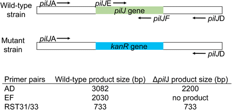
Figure 1
Figure 1
Orientation of primers for Xylella fastidiosa pilJ gene deletion. Location of binding sites for PCR primers and length of resulting PCR products for transformed XfΔpilJ strains and for wild-type control or non-transformed cells. RST31/33 are primers specific to X. fastidiosa and used for bacteria confirmation.
Figure 2
Figure 2
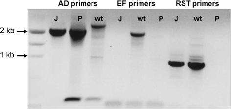
Confirmation of Xylella fastidiosa pilJ gene deletion. The pilJA/pilJD (AD) primers amplify a 3082 bp fragment from wild-type control cells (wt) or a 2200 bp fragment form the XfΔpilJ mutant (J) and deletion plasmid (P). The pilJE/pilJF (EF) primers amplify a 2030 bp band for the wild-type control strain and no band for the mutant cells or deletion plasmid. RST31/33 (RST) primers confirm that the bacteria were X. fastidiosa.
Figure 3
Figure 3
Mixture of wild-type and mutant Xylella fastidiosa strains after first isolation. The XfΔpilJ mutant confirmed in Figure 2 was stored at -80°C, streaked onto periwinkle agar plates amended with kanamycin, and the genotype assessed for 12 single colonies. Each number denotes a single colony. The pilJE/pilJF (EF) primers amplified a 2030 bp band for non-transformed X. fastidiosa and no band for the XfΔpilJ mutant. Wild-type X. fastidiosa DNA (wt) was used as a positive control for the PCR reaction, while primer reaction without template DNA represented by H2O, was used as a negative control.
Figure 4
Figure 4
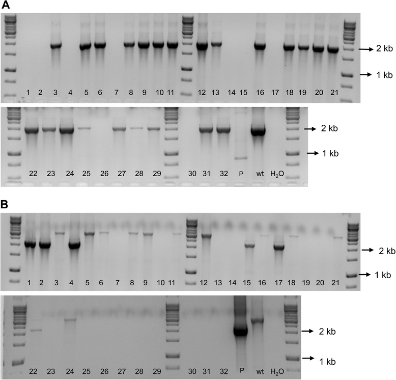
Mixture of wild-type and mutant Xylella fastidiosa strains after second isolation. The XfΔpilJ mutant confirmed in Figure 3 (isolate 12) was streaked onto periwinkle agar plates amended with kanamycin and the genotype assessed for 32 single colonies. Each number denotes a single colony. A. The pilJE/pilJF (EF) primers amplified a 2030 bp band for non-transformed bacteria and no equivalent bands for the transformed XfΔpilJ mutant strains or the deletion plasmid (P). B. The pilJA/pilJD (AD) primers amplified a 3082 bp band for non-transformed cells and a 2200 bp fragment from the XfΔpilJ strain and the deletion plasmid (P). Wild-type X. fastidiosa DNA (wt) was used as a positive control for the PCR reactions, while primer reaction without template DNA represented by H2O, was used as negative controls for each PCR reaction.
Figure 5
Figure 5
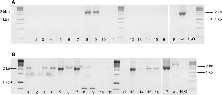
Mixture of wild-type and mutant Xylella fastidiosa strains after third isolation. The XfΔpilJ mutants confirmed in Figure 4 (isolates 4 and 17) were streaked onto PW agar plates amended with kanamycin and the genotype assessed for 16 single colonies. Each number denotes a single colony. A. The pilJE/pilJF (EF) primers amplified a 2030 bp band for non-transformed bacteria and no equivalent band for the XfΔpilJ strains or the deletion plasmid (P). B. The pilJA/pilJD (AD) primers amplified a 3082 bp band for non-transformed cells and a 2200 bp fragment from the XfΔpilJ strains and the deletion plasmid (P). Wild-type X. fastidiosa DNA (wt) was used as a positive control for the PCR reactions, while primer reaction without template DNA represented by H2O, was used as negative controls for both PCR reactions.
Figure 6
Figure 6
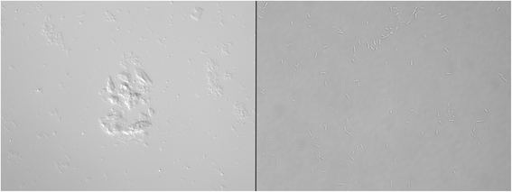
Aggregation of Xylella fastidiosa and Escherichia coli. A suspension of X. fastidiosa (left) or E. coli (right) in SCP buffer five minutes after vigorous resuspension by vortexing and pipetting. Twenty microliters was pipetted onto slides and viewed by microscopy at 40X. While E. coli cells dispersed, X. fastidiosa present as aggregates, and could not be evenly dispersed.
Figure 7
Figure 7
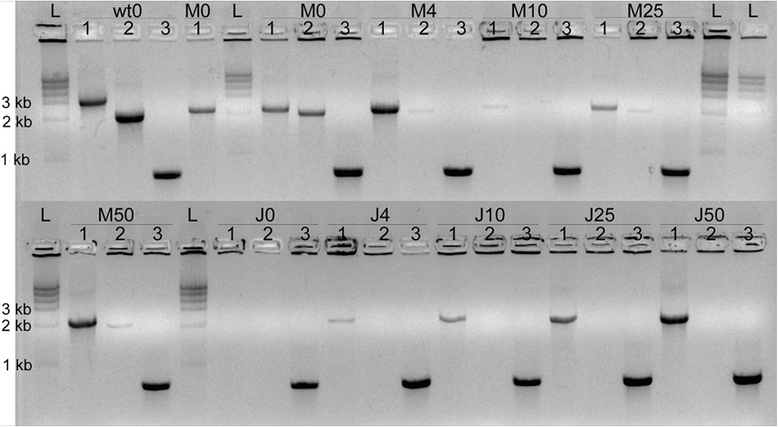
Protection of wild-type Xylella fastidiosa from antibiotic selection pressure. Wild-type X. fastidiosa (wt), XfΔpilJ mutant (J), or an equal mixture of both (M) were grown in PD2 liquid media before being plated onto agar plates with 0, 4, 10, 25, or 50 μg/mL kanamycin, and tested by PCR. The pilJA/pilJD (AD) primers amplified a 3082 bp band for wild-type cells and a 2200 bp fragment from the XfΔpilJ strain. The pilJE/pilJF (EF) primers amplified a 2030 bp band for wild-type bacteria and no equivalent bands for the XfΔpilJ mutant strains. For mixed samples, the AD primers amplified a Xf pilJ strain fragment and the EF primers amplified a wild-type band. The RST31/33 (RST) primers confirmed that the bacteria were X. fastidiosa. Wells of each cell type and kanamycin concentration condition are numbered as follows: (1) AD amplification, (2) EF amplification, and (3) RST amplification.

Similar articles

References

    1. Chatterjee S, Almeida RP, Lindow S. Living in two worlds: the plant and insect lifestyles of Xylella fastidiosa. Annu Rev Phytopatho. 2008;46:243–71. doi: 10.1146/annurev.phyto.45.062806.094342. - DOI - PubMed
    1. Meng Y, Li Y, Galvani CD, Hao G, Turner JN, Burr TJ, et al. Upstream migration of Xylella fastidiosa via pilus-driven twitching motility. J Bacterio. 2005;187(16):5560–7. doi: 10.1128/JB.187.16.5560-5567.2005. - DOI - PMC - PubMed
    1. Mattick JS. Type IV, pili and twitching motility. Annu Rev Microbio. 2002;56:289–314. doi: 10.1146/annurev.micro.56.012302.160938. - DOI - PubMed
    1. Feil H, Feil WS, Lindow SE. Contribution of fimbrial and afimbrial adhesins of Xylella fastidiosa to attachment to surfaces and virulence to grape. Phytopathology. 2007;97(3):318–24. doi: 10.1094/PHYTO-97-3-0318. - DOI - PubMed
    1. De La Fuente L, Burr TJ, Hoch HC. Autoaggregation of Xylella fastidiosa cells is influenced by type I and type IV pili. Appl Environ Microbiol. 2008;74(17):5579–82. doi: 10.1128/AEM.00995-08. - DOI - PMC - PubMed

Publication types

LinkOut - more resources