. 2015 Apr 16:5:9649.
doi: 10.1038/srep09649.

Molecular basis for mid-region amyloid-β capture by leading Alzheimer's disease immunotherapies

Affiliations

Molecular basis for mid-region amyloid-β capture by leading Alzheimer's disease immunotherapies

Gabriela A N Crespi et al. Sci Rep. .

Abstract

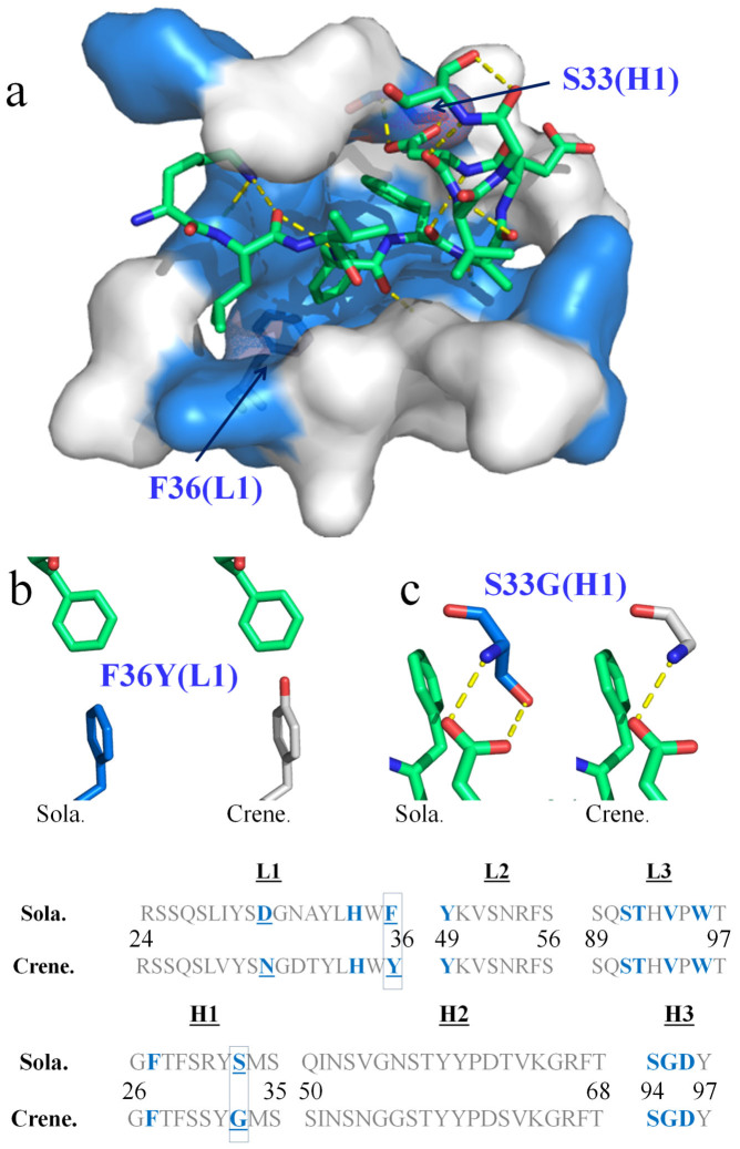
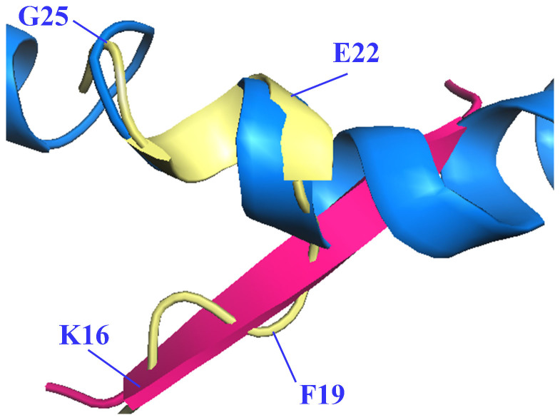
Solanezumab (Eli Lilly) and crenezumab (Genentech) are the leading clinical antibodies targeting Amyloid-β (Aβ) to be tested in multiple Phase III clinical trials for the prevention of Alzheimer's disease in at-risk individuals. Aβ capture by these clinical antibodies is explained here with the first reported mid-region Aβ-anti-Aβ complex crystal structure. Solanezumab accommodates a large Aβ epitope (960 Å(2) buried interface over residues 16 to 26) that forms extensive contacts and hydrogen bonds to the antibody, largely via main-chain Aβ atoms and a deeply buried Phe19-Phe20 dipeptide core. The conformation of Aβ captured is an intermediate between observed sheet and helical forms with intramolecular hydrogen bonds stabilising residues 20-26 in a helical conformation. Remarkably, Aβ-binding residues are almost perfectly conserved in crenezumab. The structure explains the observed shared cross reactivity of solanezumab and crenezumab with proteins abundant in plasma that exhibit this Phe-Phe dipeptide.


Figures

Figure 1
Figure 1. Structure of the mid-region of the Aβ peptide bound to solanezumab.
Both panels show Aβ nestled in the surface of the Fab CDRs. Solanezumab is shown as a transparent surface, light blue (light chain) and darker blue (heavy chain). (a) Both copies of the peptide in the asymmetric unit are shown in lime and yellow sticks. Overall conformation of Aβ as recognised by solanezumab; amino acids of the Aβ epitope are labelled. (b) Helical conformation adopted by Aβ residues C-terminal to the buried Phe19-Phe20 dipeptide. The view is taken 90 degrees rotation about the Y-axis from that shown in panel (a).
Figure 2
Figure 2. The clinical antibodies solanezumab and crenezumab recognise Aβ in almost identical fashion.
(a) The Aβ peptide (lime sticks) is shown bound to solanezumab through its CDRs. The CDRs are represented as a surface with Aβ-contacting residues coloured blue. Polar contacts are exhibited as yellow dashed lines. The CDR sequences (L1, L2 and L3 from the light chain, and H1, H2 and H3 from the heavy chain) of solanezumab (sola.) and the clinical immunotherapy crenezumab (crene.) are shown at the bottom of the figure. Each CDR loop in solanezumab is the same length as its counterpart in crenezumab. Antibody residues that contact Aβ in the solanezumab-Aβ complex structure, and the corresponding residues in crenezumab, are coloured blue. The crenezumab-Aβ complex structure was derived by homology modelling from the solanezumab-Aβ complex crystal structure (see text). Only two of those contacting residues are not conserved: namely, Sola. residues Phe36(L1) and Ser33(H1). These are labelled in (a) and their local environments are highlighted respectively in (b) and (c).
Figure 3
Figure 3. Different conformations of the mid-region of the Aβ peptide.
Aβ structure as recognised by solanezumab (PDB id 4XXD is shown as light yellow cartoon with every third Cα labelled), superpositioned across residues KLVF derived from β-sheet crystallographic structures (PDB id: 4NTR (pink)), and across residues FAEDVGS with the HFIP-induced solution state helical Aβ structure (PDB id: 1Z0Q, marine blue).

References

    1. Herrmann A. & Spires-Jones T. Clearing the way for tau immunotherapy in Alzheimer's disease. J. Neurochem. 132, 1–4 (2015). - PubMed
    1. Hardy J. et al. Pathways to Alzheimer's disease. J. Intern. Med. 275, 296–303 (2014). - PubMed
    1. Adolfsson O. et al. An effector-reduced anti-β-amyloid (Aβ) antibody with unique Aβ binding properties promotes neuroprotection and glial engulfment of Aβ. J. Neurosci. 32, 9677–9689 (2012). - PMC - PubMed
    1. Legleiter J. et al. Effect of different anti-Abeta antibodies on Abeta fibrillogenesis as assessed by atomic force microscopy. J. Mol. Biol. 335, 997–1006 (2004). - PubMed
    1. Choi S. H. et al. A three-dimensional human neural cell culture model of Alzheimer's disease. Nature 515, 274–278 (2014). - PMC - PubMed

Publication types

MeSH terms

Associated data