Review
. 2015 Oct:34:125-32.
doi: 10.1016/j.conb.2015.03.010. Epub 2015 Apr 14.

Visual circuits in flies: beginning to see the whole picture

Affiliations
Review

Visual circuits in flies: beginning to see the whole picture

Rudy Behnia et al. Curr Opin Neurobiol. 2015 Oct.

Abstract

Sensory signals are processed in the brain by dedicated neuronal circuits to form perceptions used to guide behavior. Drosophila, with its compact brain, amenability to genetic manipulations and sophisticated behaviors has emerged as a powerful model for investigating the neuronal circuits responsible for sensory perception. Vision in particular has been examined in detail. Light is detected in the eye by photoreceptors, specialized neurons containing light sensing Rhodopsin proteins. These photoreceptor signals are relayed to the optic lobes where they are processed to gain perceptions about different properties of the visual scene. In this review we describe recent advances in the characterization of neuronal circuits underlying four visual modalities in the fly brain: motion vision, phototaxis, color and polarized light vision.


Figures

Figure 1
Figure 1. The eye and the optic lobe of adult Drosophila
A A single ommatidium contains eight photoreceptors, six outers R1-6 (grey) and two inners R7-8 (colors). Outers express the Rh1 opsin, R7s express either Rh3 or Rh4 while R8s express Rh5 or Rh6. Four types of ommatida are found in the eye. ‘Pale’ and ‘yellow’ ommatidia are distributed stochastically in the main part of the eye. In the dorsal third, pale and specialized dorsal third yellow subtypes are found (not shown). In one to two rows of ommatidia in the dorsal rim are (DRA) of the retina, the remaining DRA subtypes are found. B Stochastic distribution of Rh5 (blue) and Rh6 (green) expressing R8s in the main part of the eye. C Normalized spectral preference curves of the different rhodopsins expressed in the eye of the fly. Rh1 shows broad spectral sensitivity peaking in both the blue and the UV due to the presence of a sensitizing pigment (Modified from ) D Photoreceptors project to the optic lobe. Outer photoreceptors send their axons to the lamina while R7/R8 photoreceptors send theirs to the medulla. The lobula is involved in spectral preference, color and polarized light vision. The lobula plate is a center for motion detection.
Figure 2
Figure 2. Motion vision in the Drosophila optic lobe
A. The Hassentein and Reichard Correlator (HRC) relies on differential temporal filtering of two spatially separated input channels, delaying one signal with respect to the other. Motion from left to right in this case causes these delayed and non-delayed luminance signals to arrive simultaneously at a subsequent processing step where they are multiplied and amplified (multiplication) as a motion signal. Motion in the opposite direction where the delay separates the signals in time, leads to a null output. B The subtraction of the output of a correlator from that of a mirror-imaged correlator produces responses that have different signs for opposite directions. (A and B modified from ) C Two pathways lead from photoreceptors in the eyes to LPTCs. In the moving light-edge-specific pathway, L1 neurons, postsynaptic to photoreceptors, act as inputs, while direction selective T4 neurons, presynaptic to LPTCs, act as outputs. L2 and T5 have equivalent roles in the pathway that detects moving dark edges. Medulla neurons Mi1/Tm3, Tm1/Tm2 have been proposed to be the delayed and the non-delayed lines of an HRC for moving light edges respectively. The dendrites of T4/T5 neurons define potential sites for HRC signal multiplication in these two pathways. D T4 cells respond preferentially to moving bright edges, T5 cells respond to moving dark edges. Dendrites responding to different cardinal directions are located to fours different layers of the lobula plate.
Figure 3
Figure 3. Spectral preference, color vision and polarized light vision in the Drosophila optic lobe
A Dm8 and Tm5c neurons are necessary for UV spectral preference. Each Dm8 gets inhibitory inputs from approximately 16 R7s. The information from Dm8 is in then passed on through excitatory synapses to Tm5c which projects to the lobula (modified from ). B Color vision cannot be achieved by single photoreceptors that respond to light over a broad range of wavelengths. Instead, it necessitates the comparison of the output of photoreceptors with different spectral sensitivity (top, showing hypothetical blue and green Rhodopsin-expressing photoreceptor sensitivity). Neural response of a color opponent cell which gets excitatory input from the blue sensitive photoreceptor and inhibitory input from the green sensitive photoreceptor shown above. The response of this neuron increases for wavelengths below the x-axis crossing point and decreases above it, no mater the intensity. C Light is an electromagnetic wave. Its electric and magnetic fields vibrate in planes perpendicular to each other and to the direction of wave travel (black arrow). When all the electric field vectors lie in a plane, the wave is linearly polarized. The orientation of this plane is the direction of the e-vector of polarization. D Pattern of polarized light of the sky. E-vectors are arranged along concentric circles around the sun shown in yellow (B and C from ).

References

    1. Johnston RJ. Lessons about terminal differentiation from the specification of color-detecting photoreceptors in the Drosophila retina. Annals of the New York Academy of Sciences. 2013;1293:33–44. - PubMed
    1. Heisenberg M, Buchner E. The role of retinula cell types in visual behavior of Drosophila melanogaster. Journal of comparative physiology. 1977;117:127–162.
    1. Schnaitmann C, Garbers C, Wachtler T, Tanimoto H. Color Discrimination with Broadband Photoreceptors. Current Biology. 2013;23:2375–2382. - PubMed
    1. Melnattur KV, et al. Multiple Redundant Medulla Projection Neurons Mediate Color Vision in Drosophila. Journal of neurogenetics. 2014:1–15. - PMC - PubMed
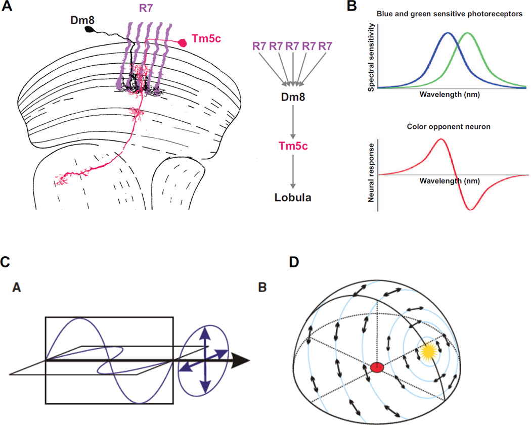
    1. Wernet MF, et al. Genetic Dissection Reveals Two Separate Retinal Substrates for Polarization Vision in Drosophila. Current Biology. 2012;22:12–20. The authors of this article describe a novel behavioral paradigm to dissect the neuronal circuits for polarization vision. They show that flies exhibit detection of ventral as well as dorsal polarization and dissect the contribution of different photoreceptor types in the eye of the fly to these two polarization driven behaviors.

Publication types

LinkOut - more resources