. 2015 Jun 15;118(12):1525-32.
doi: 10.1152/japplphysiol.00934.2014.

Nasal high flow clears anatomical dead space in upper airway models

Nasal high flow clears anatomical dead space in upper airway models

Winfried Möller et al. J Appl Physiol (1985). .

Abstract

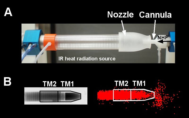
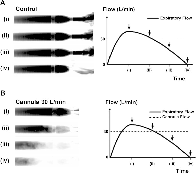
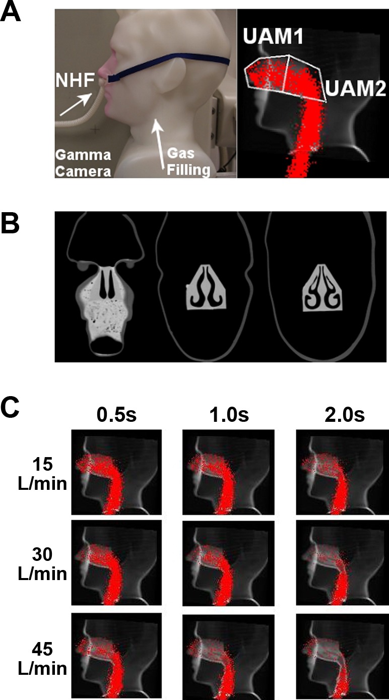
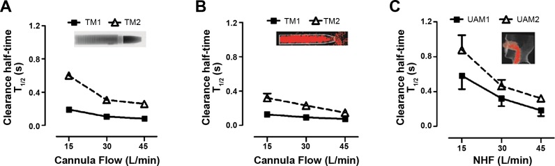
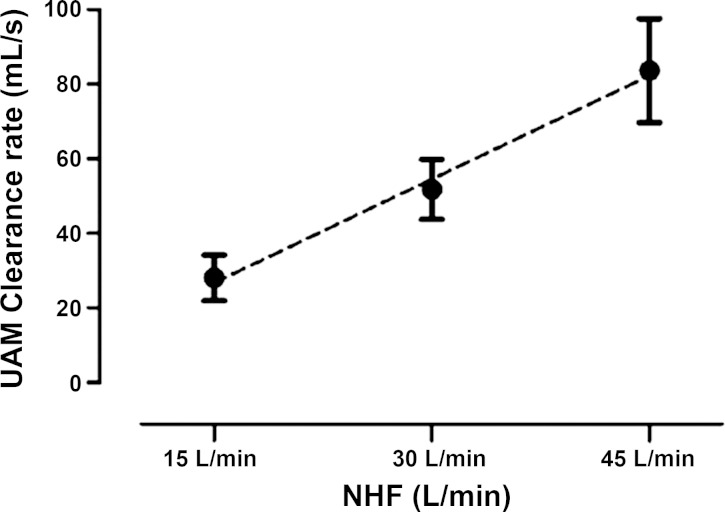
Recent studies showed that nasal high flow (NHF) with or without supplemental oxygen can assist ventilation of patients with chronic respiratory and sleep disorders. The hypothesis of this study was to test whether NHF can clear dead space in two different models of the upper nasal airways. The first was a simple tube model consisting of a nozzle to simulate the nasal valve area, connected to a cylindrical tube to simulate the nasal cavity. The second was a more complex anatomically representative upper airway model, constructed from segmented CT-scan images of a healthy volunteer. After filling the models with tracer gases, NHF was delivered at rates of 15, 30, and 45 l/min. The tracer gas clearance was determined using dynamic infrared CO2 spectroscopy and 81mKr-gas radioactive gamma camera imaging. There was a similar tracer-gas clearance characteristic in the tube model and the upper airway model: clearance half-times were below 1.0 s and decreased with increasing NHF rates. For both models, the anterior compartments demonstrated faster clearance levels (half-times < 0.5 s) and the posterior sections showed slower clearance (half-times < 1.0 s). Both imaging methods showed similar flow-dependent tracer-gas clearance in the models. For the anatomically based model, there was complete tracer-gas removal from the nasal cavities within 1.0 s. The level of clearance in the nasal cavities increased by 1.8 ml/s for every 1.0 l/min increase in the rate of NHF. The study has demonstrated the fast-occurring clearance of nasal cavities by NHF therapy, which is capable of reducing of dead space rebreathing.


Figures

Fig. 1.
Fig. 1.
A: upper airway tube model (TM) made from a sapphire tube and a sodium chloride (NaCl) nozzle with a cannula inserted into the nozzle in front of the IR-heat radiation source (blackbody). Also shown are the pressure ports and the pneumotachographs to monitor pressure and flow within the tube and the cannula. The cannula flow rates [nasal high flow (NHF)] were delivered into the nozzle at 15, 30, and 45 l/min. B: an infrared absorption image (left) and a gamma camera image (right) show the filling stage of the model with CO2 and 81mKr gas before air was flushed into the cannula. Anterior (TM1) and posterior (TM2) ROIs were defined for data analysis.
Fig. 2.
Fig. 2.
Infrared absorption images of expiratory flow through a tube model (TM) of upper airways demonstrate rebreathing from dead space. The images show four stages of filling of the model with exhaled CO2 at (i) peak expiratory flow, (ii) expiratory flow 30 l/min, (iii) expiratory flow 15 l/min, and (iv) end of expiration. A: control demonstrates filling of the TM during the expiration phase without NHF from a cannula. At the beginning of inspiration all gas from the TM will be rebreathed into the lungs. B: NHF from the cannula purges the expired CO2-rich gas from the model and replaces it with fresh air. This results in a reduction of CO2 rebreathing. Breathing through the model demonstrates that the replacement of expired gas with air starts before the end of expiration and that the static conditions used in the experiments led to an underestimation of the speed of dead-space clearance during respiration.
Fig. 3.
Fig. 3.
A, left: standard image of the upper airway model (UAM) showing the setup of the cannula interface (left panel) in the nostrils. A, right: the same image overlaid with the outlines of the anterior (UAM1) and posterior (UAM2) ROIs in the nasal cavities, and data from the planar gamma camera when the UAM was filled from the trachea end with 81mKr-gas. B: coronary CT scans of the model, illustrating the complex internal anatomical structure in the UAM. C: lateral gamma camera images of 81mKr-gas filling of UAM superimposed onto a sagittal CT of the UAM. Series of images illustrate the tracer-gas clearance at time points 0.5, 1.0, and 2.0 s using NHF rates 15, 30, and 45 l/min.
Fig. 4.
Fig. 4.
Comparison of clearance profiles during flow rates of 15, 30, and 45 l/min from a custom-made cannula in the TM and a standard cannula interface in the UAM model using comparable ROIs. A: clearance half-time (T1/2) in the TM with CO2-gas MWIR imaging experiments. B: clearance half-time (T1/2) in the TM with 81mKr-gas gamma imaging experiments. C: clearance half-time (T1/2) in the UAM with 81mKr-gas gamma imaging experiments. The clearance profiles are similar in all three experiments. Anterior ROIs (TM1 and UAM1) are cleared faster than posterior ROIs (TM2 and UAM2). Clearance in posterior ROIs is more flow dependent than in anterior ROIs.
Fig. 5.
Fig. 5.
Clearance rates in nasal cavities (total volume 55 ml) of the upper airway model (UAM) at NHF rates of 15, 30, and 45 l/min, calculated from the clearance half-times and corresponding volumes of UAM1 and UAM2 ROIs. The clearance rate linearly rises with an increase of NHF. The graph shows that in the static experimental setup NHF of 30 l/min clears the total volume of the nasal cavity within 1 s.

References

    1. Ari A, Harwood R, Sheard M, Dailey P, Fink JB. In vitro comparison of heliox and oxygen in aerosol delivery using pediatric high flow nasal cannula. Pediatr Pulmonol 46: 795–801, 2011. - PubMed
    1. Bott J, Carroll MP, Conway JH, Keilty SEJ, Ward EM, Brown AM, Paul EA, Elliott MW, Godfrey RC, Wedzicha JA, Moxham J. Randomised controlled trial of nasal ventilation in acute ventilatory failure due to chronic obstructive airways disease. Lancet 341: 1555–1557, 1993. - PubMed
    1. Bräunlich J, Beyer D, Mai D, Hammerschmidt S, Seyfarth HJ, Wirtz H. Effects of nasal high flow on ventilation in volunteers, COPD and idiopathic pulmonary fibrosis patients. Respiration 85: 319–325, 2013. - PubMed
    1. Dhand R. Aerosol therapy in patients receiving noninvasive positive pressure ventilation. J Aerosol Med Pulm Drug Deliv 25: 63–78, 2011. - PubMed
    1. Dysart K, Miller TL, Wolfson MR, Shaffer TH. Research in high flow therapy: mechanisms of action. Respir Med 103: 1400–1405, 2009. - PubMed

Publication types

LinkOut - more resources