Review
. 2015 Sep;1851(9):1156-85.
doi: 10.1016/j.bbalip.2015.04.002. Epub 2015 Apr 13.

As the fat flies: The dynamic lipid droplets of Drosophila embryos

Affiliations
Review

As the fat flies: The dynamic lipid droplets of Drosophila embryos

Michael A Welte. Biochim Biophys Acta. 2015 Sep.

Abstract

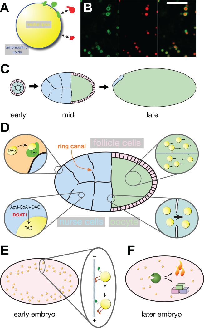
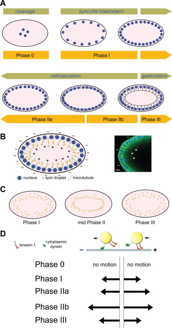
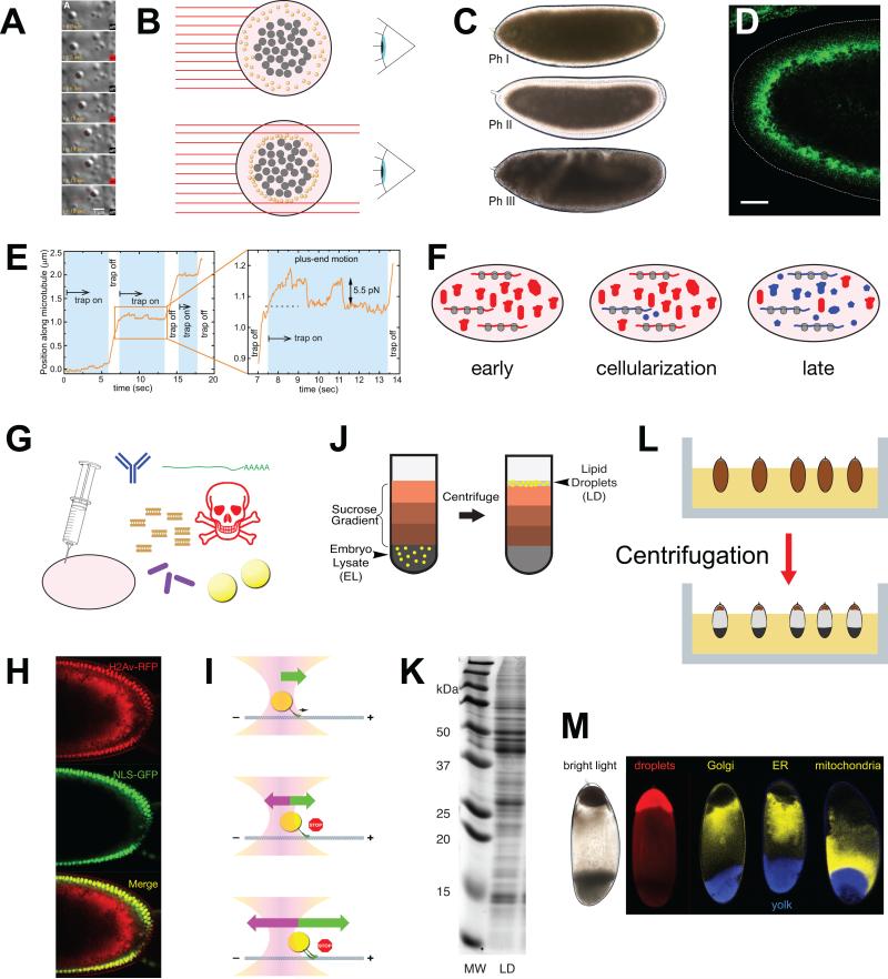
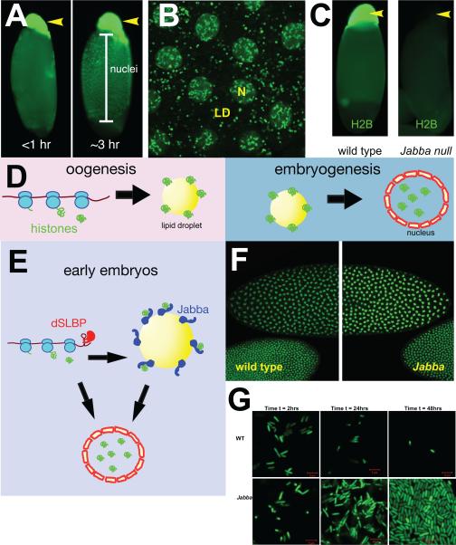
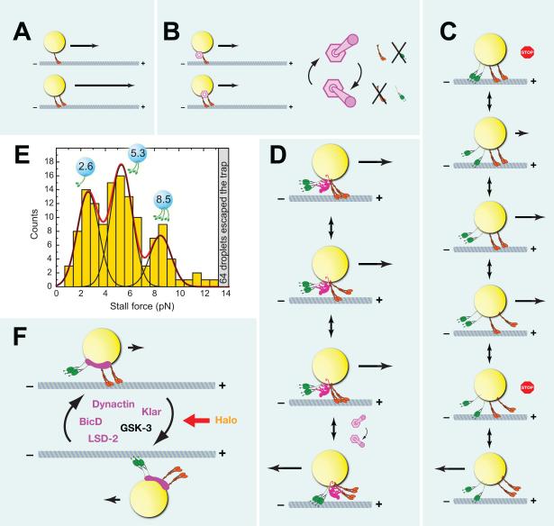
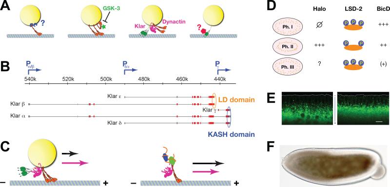
Research into lipid droplets is rapidly expanding, and new cellular and organismal roles for these lipid-storage organelles are continually being discovered. The early Drosophila embryo is particularly well suited for addressing certain questions in lipid-droplet biology and combines technical advantages with unique biological phenomena. This review summarizes key features of this experimental system and the techniques available to study it, in order to make it accessible to researchers outside this field. It then describes the two topics most heavily studied in this system, lipid-droplet motility and protein sequestration on droplets, discusses what is known about the molecular players involved, points to open questions, and compares the results from Drosophila embryo studies to what it is known about lipid droplets in other systems.

Keywords: Drosophila embryo; Lipid droplet; Microtubule motors; Protein sequestration.


Figures

Figure 1
Figure 1. The life cycle of Drosophila embryonic lipid droplets
(A) Structure of lipid droplets: A core of neutral lipids (triglycerides (TAG), sterol esters, retinol esters) is surrounded by a monolayer of amphipathic lipids (such as phosphoglycerides and sterols). Proteins can be stably embedded (green) in this monolayer or reversibly bound (red) to other proteins or lipid head groups. (B) Colocalization of a GFP fusion (green) that targets to lipid droplets (GFP-LD [56]) and neutral lipids (red, detected with the dye Nile Red). The fusion protein surrounds a core of neutral lipids. Note that this particular fusion protein labels only a subset of lipid droplets. Scale bar = 5 μm. Image modified from [56]. (C) Overview of oogenesis (for color code of cell types, see D). In early egg chambers, the 16-daughter cells of the germ-line cytoblast (the future nurse cells and oocytes) are surrounded by a layer of somatic follicle cells. In mid-stage egg chambers, nurse cells supply the growing oocyte with nutrients, proteins and RNAs; follicle cells have migrated to cover the oocyte. In late stages, nurse cells have transferred most of their contents to the oocyte and have undergone apoptosis. The oocyte is surrounded by an eggshell (not shown) that was produced by the follicle cells. The various stages are not drawn to scale; by the end of oogenesis the oocyte volume has increased more than a hundred fold. (D) Lipid droplets in mid-stage egg chambers. One of the ring canals connecting nurse cells and oocyte is indicated. Top left: lipophorin particles (diacylglycerol (DAG) rich components of the hemolymph) are taken up by nurse cells via lipophorin receptors (Lpr). Bottom left: in the nurse cells, DGAT1 converts acyl-CoA and DAG into TAG, which contributes to the growth of the LD core. Bottom right: cytoplasmic streaming transports lipid droplets from nurse cells through ring canals into the oocyte. Top right: in the oocyte cytoplasm, most lipid droplets move passively by cytoplasmic streaming; a subset is actively transported by motors along microtubules. (E) In early embryos, lipid droplets move bidirectionally along microtubules. (F) Later in embryogenesis, lipid droplets are thought to be broken down to generate energy (via oxidative phosphorylation) and building blocks (for biomass production).
Figure 2
Figure 2. Lipid droplet motility in the early embryos
(A) Overview of early embryogenesis. During cleavage stages nuclei divide in the embryo interior. By syncytial blastoderm (nuclear cycle 10-13), a subset of nuclei has reached the surface and continues to undergo mitosis. During cellularization (nuclear cycle 14), plasma membranes grow in between the nuclei, converting the syncytial embryo with thousands of nuclei into a monolayer of cells with one nucleus per cell. Gastrulation movements start shortly thereafter. The orange bars represent the extent of the phases of lipid-droplet transport relative to other morphological events. Embryos are drawn schematically and not to scale; in reality, there are some ~6000 nuclei present at the periphery at cellularization. In addition, from syncytial blastoderm onward, the center of the embryo also contains polyploidy yolk nuclei; for clarity, they have been omitted from the cartoon. (B) Left: Schematic representation of the distribution of nuclei, lipid droplets, and microtubules in early embryos (not drawn to scale). Nuclei are present close to the embryo surface, all around the embryo. Microtubules are oriented radially, with their minus ends close to the surface and their plus ends pointing into the embryo interior. Right: Micrograph of parts of an early embryo, depicting DNA (blue, to highlight nuclei) and microtubules (green). Microtubule polarity (+ and – ends) is indicated in a few instances. Scale bar = 10 μm. Image modified from [76]. (C) Schematic representation of the global distribution of lipid droplets during early embryogenesis (not drawn to scale). In Phase I and III, droplets are found all over the peripheral cytoplasm. In Phase II, droplets relocate inward and accumulate around the central yolk (not shown, but see Fig. 3B). (D) Cartoon of lipid-droplet motion: Droplets move back and forth along microtubules, powered by the plus-end motor kinesin-1 and the minus-end motor cytoplasmic dynein. The arrows show the relative lengths of movements in the minus- and plus-end directions in various phases (based largely on the measurements in [83], but see also [86] and [57]; the exact run-length values vary with the tracking method employed; the figure summarizes the relative balance of motion in the two directions).
Figure 3
Figure 3. Methods to study Drosophila embryonic droplets
(A) Lipid-droplet motion visualized by differential interference contrast (DIC) microscopy and manipulated by an optical tweezer/trap. Time in seconds is indicated as well as whether the laser (centered on the droplet) is turned on or off. Red line shows the position of the center of the lipid droplet tracked over time. The droplet proceeded steadily along a linear path (inferred to be a microtubule) and its progress was impeded by the optical trap. For details of the tracking and laser trap analysis, see E. (Image from [59]). (B) How lipid droplet distribution affects embryo transparency. Shown is an embryo cross-section perpendicular to the long axis of the embryo. Light (red lines) from a source on the left passes through the embryo and is collected by an observer on the right. Yolk granules (gray) and lipid droplets (yellow) scatter light and thus prevent light from passing through the cytoplasm. In the top embryo, lipid droplets are spread out all over the periphery and block light evenly. In the bottom embryo, lipid droplets have moved away from the periphery and are accumulated around the central yolk; hence, light can pass through the periphery. (C) Changes in lipid-droplet distribution cause altered embryo transparency. In Phase I and III, the embryo periphery is opaque because the abundant lipid droplets scatter light. In Phase II, the periphery is transparent because it is depleted of lipid droplets. Image from [76]. (D) Jabba immunostaining (green) to highlight lipid droplets. The dotted line outlines the embryo surface. In this Phase II embryo, the droplets have accumulated basally, clustering around the central yolk. Scale bar = 25 μm. Image modified from [16]. (E) Movement of a lipid droplet along a microtubule as a function of time, in the presence or absence of an opposing force from an optical trap. This image is from [59] and represents the quantitation of the experiments shown in (A). The enlarged portion shows that the droplet stalls when the trap is switched on and then drops to the trap center. Another movement attempt again results in a stall. When the trap is switched off, the motors are able to continue to move the droplet. The distance at which the stall occurred is a measure for the force generated by the motor(s) moving the droplet, in this case ~5.5 pN. (F) Early embryos contain proteins directly inherited from the mother (red blobs) as well as proteins generated in the embryo from translation of maternal messaged (red). By cellularization, the zygote has started transcribing its own genes, generating its own messages and proteins (shown in blue). Later in embryogenesis, translation is driven by zygotic messages and most, but not all, proteins are the product of zygotic transcription. (G) By microinjection, various substances can be introduced into embryos, including antibodies, mRNAs, dsRNAs for RNA interference, inhibitors, bacteria, and lipid droplets. (H) Transplantation of H2Av-RFP covered lipid droplets into recipient embryos in which nuclei are marked by NLS-GFP. Merged image reveals the transplanted lipid droplets in red; some of the droplet-bound H2Av-RFP was released from droplets and was transferred into nuclei. Image from [15]. (I) Principles of optical trapping. Top: Lipid droplet in the center of an optical trap (optical tweezer). It is propelled along the microtubule by the force generated by the microtubule motor. The trap does not yet exert any force on the droplet. Middle: Once the droplet is displaced from the laser center, the trap exerts a force pulling the droplet back towards the center. At some point, the force from laser and motor are balanced, resulting in stalled motion. Bottom: If the droplet is pulled by multiple motors, this force balance occurs at a distance further from the center of the laser. (J) Schematic representation of lipid-droplet purification by floatation. Embryo lysate in high-density buffer is overlaid by sucrose solutions of increasingly lower density. After centrifugation, lipid droplets can be recovered at the very top of the gradient. Image from [16]. (K) Protein content of lipid-droplet fraction after sucrose gradient. Proteins from droplet fraction (LD) were analyzed by SDS PAGE. MW = molecular weight markers. Image from [16]. (L) Schematic depiction of in-vivo centrifugation of embryos. Embryos before cellularization are embedded in agar to keep them in a fixed orientation (top). After centrifugation, the contents of each embryo are separated by density (bottom). Image from [127]. (M) Separation of organelles by in-vivo centrifugation. Living embryos were centrifuged as in (L), which results in distinct stratification visible by bright-field microscopy (left). Distribution of various organelles was detected by fluorescence microscopy. Image originally from [15], as modified in [127].
Figure 4
Figure 4. Histone sequestration on droplets
(A) Centrifuged embryos stained for histone H2Av. In Phase 0 embryos, H2Av signal is almost exclusively associated with the lipid-droplet layer (yellow arrow). By Phase II, H2Av signal is found both on the droplet layer and in nuclei. Image modified from [16]. (B) In Phase I embryos, H2Av-GFP (green) is present in nuclei (large blobs, N) and on lipid droplets (small rings in the cytoplasm, LD). Image courtesy of Zhihuan Li. (C) In-vivo centrifugation demonstrates that histone H2B is present on lipid droplets in a Jabba-dependent manner. Both panels show Phase 0 embryos after centrifugation; lipid-droplet layers are indicated by yellow arrows. Left: wild-type embryo stained for H2B (H2B is highly enriched on the droplet layer). Right: Jabba mutant embryo stained for H2B (H2B is absent from the droplet layer). Image modified from [16]. (D) Storage of histones on lipid droplets allows temporal uncoupling of histone production and usage: During oogenesis, newly synthesized histones are sequestered on lipid droplets. The sequestered histones are released during embryogenesis and are relocated to the nucleus to package chromatin. (E) Histone dynamics in early embryos. Early embryos contain both histone mRNAs and histone proteins provided from the mother. The translation of the messages for canonical histones is regulated by the Drosophila stem loop binding protein (dSLBP). Maternal histone proteins are stored on lipid droplets via binding to Jabba. Both newly translated histones and droplet-stored histones contribute to chromatin assembly in the nucleus. In addition, excess newly synthesized histone can be sequestered on droplets, thus buffering the histone supply. (F) Wild-type and Jabba mutant embryos stained for H2Av. At certain stages, Jabba mutant embryos display overaccumulation of H2Av in their nuclei. Image modified from [75]. (G) Wild-type and Jabba mutant embryos injected with E. coli expressing GFP. While over time the bacterial population declines in the wild-type embryos, it dramatically increases in Jabba mutants. Image from [19].
Figure 5
Figure 5. The motors driving lipid droplet motion
(A) Motor behavior in vitro: Cargo moved by two motors can travel for considerable longer distances than cargo moved by a single motor. Arrows indicate distance traveled. (B) Proposed “switch” model for lipid-droplet transport in vivo. Left: a switch mechanism terminates motion independent of motor number; both cargoes move the same distance. Arrows indicate distance traveled. Right: For bidirectional transport, the switch toggles between two states: “kinesin-1 ON, cytoplasmic dynein OFF” and “kinesin-1 OFF, cytoplasmic dynein ON” (right). (C) Bidirectional transport as a result of a tug-of-war between opposing motors. Arrows indicate travel velocity. If the numbers and forces of opposing motors are well balanced, cargoes will frequently be stalled, in severe motor competition (panel 1). As motors attach and detach stochastically, motor imbalance will arise that allows slow motion in a particular direction (panel 2). If motors under load release more readily, this imbalance will quickly resolve itself into only motors for one direction being actively engaged on the microtubule (panel 3). Stochastic binding/release of motors will re-establish the paused state and can even result in reversal of direction (panels 4, 5, 6). (D) Bidirectional transport as the result of the still hypothetical coordination machinery (pink): The coordination machinery keeps cytoplasmic dynein off (possibly by sterically preventing binding to the track) while the opposing kinesin-1 motors are on. Once the switch is triggered, the coordination machinery turns kinesin-1 off and simultaneously makes cytoplasmic dynein active. (E) Stall force measurements for plus-end directed lipid droplets in Drosophila embryos show peaks at multiples of ~2.6 pN. This pattern indicates the action of 1, 2, or 3 kinesins per droplet. Image from [59]. (F) Factors known to regulate lipid-droplet motion. Lipid droplets constantly switch between motion dominated by the plus-end motor (top) and motion dominated by the minus-end motor (bottom). The pink blob represents a hypothesized switching complex. Dynactin and Klar have been proposed to act as integral parts of the switch mechanism involved. BicD, GSK-3, and LSD-2 also affect the distance traveled in one or both directions, and thus may be involved in flipping the switch. Halo acts as transacting signal that mediates the temporal pattern of switching frequency. Klar, LSD-2, Dynactin, and BicD are localized to lipid droplets and may be part of the switching complex. Whether GSK-3 or Halo are physically present on the droplets is unknown.
Figure 6
Figure 6. Regulators of lipid-droplet transport
(A) Proposed models of force regulation during lipid-droplet transport (from left to right): 1) Motor number per droplet might be controlled by the availability of docking sites or the number of motors available for docking. The cargo adaptor for motors on droplets is not yet known [76]. 2) The activity of motors might be controlled after docking; GSK-3 has been proposed to restrict the activity of docked kinesin-1 [85]. 3) Motor coordinators may allow full force production by keeping opposite-polarity motors inactive, as proposed for dynactin and Klar [57, 84]. 4) Cytoplasmic dynein can exist in low- and high-force states [312], and these states can be controlled by transacting factors [214]. So force production by cytoplasmic dynein on lipid droplets might be regulated in vivo, an idea that has not yet been tested. (B) The complex klar locus encodes five different protein isoforms, α, β, γ, δ and ε. Promoters are indicated by blue arrows, non-coding exons by gray bars, and coding exons by red/orange/blue bars. LD domain is shown in orange, KASH domain in blue. Map modified after [266]. (C) Comparison of the effect of Klar on lipid droplet (left) and mRNA (right) transport. Arrows symbolize run lengths in the presence (pink) or absence (black) of Klar. Presence of Klar increases plus-end travel lengths for lipid droplets, but reduces them for RNP particles [57, 235]. (D) Differences in Halo, LSD-2, and BicD proteins between phases of droplet transport. Halo is absent in Phase I and expressed in Phase II; its status in Phase III is unknown, but circumstantial evidence and halo's mRNA expression pattern has led to the proposal that Halo is degraded by this time [79, 86]. LSD-2 is highly phosphorylated in Phase I and III, but less so in Phase II [87]. Droplet levels of BicD protein drop progressively from Phase I to II to III [83]. (E) Halo acts as a directionality determinant for transport. GFP-labeled lipid droplets in late Phase IIa embryos in which Halo is either expressed (left) or missing (right). The dotted line outlines the embryo surface. In the presence of Halo, net transport is plus-end directed (inward); in the absence of Halo, net transport is minus-end direction (outward). Scale bar = 10 μm. Image modified from [56]. (F) Acute effect of Halo on lipid-droplet distribution and embryo transparency. Bright-field image of a Phase IIa embryo mutant for Halo in which in-vitro generated halo mRNA was injected on the right. In the left half of the embryo, Halo activity was absent and lipid droplets are spread throughout the periphery, resulting in a broad brown “halo” around the central yolk. In the right half of the embryo, Halo activity was present, and lipid droplets accumulated around the central yolk (as a narrow dark band), leaving the periphery depleted of droplets. As a result, the peripheral cytoplasm is transparent. Image from [86].

References

    1. Martin S, Parton RG. Lipid droplets: a unified view of a dynamic organelle. Nat Rev Mol Cell Biol. 2006;7:373–378. - PubMed
    1. Walther TC, Farese RV., Jr. Lipid droplets and cellular lipid metabolism. Annu Rev Biochem. 2012;81:687–714. - PMC - PubMed
    1. Igal RA, Coleman RA. Neutral lipid storage disease: a genetic disorder with abnormalities in the regulation of phospholipid metabolism. J Lipid Res. 1998;39:31–43. - PubMed
    1. Greenberg AS, Coleman RA, Kraemer FB, McManaman JL, Obin MS, Puri V, Yan QW, Miyoshi H, Mashek DG. The role of lipid droplets in metabolic disease in rodents and humans. J Clin Invest. 2011;121:2102–2110. - PMC - PubMed
    1. Paul A, Chan L, Bickel PE. The PAT family of lipid droplet proteins in heart and vascular cells. Curr Hypertens Rep. 2008;10:461–466. - PMC - PubMed

Publication types

MeSH terms

Substances

LinkOut - more resources