. 2015 May 29;290(22):13948-57.
doi: 10.1074/jbc.M115.655043. Epub 2015 Apr 16.

A Point Mutation in the Ubiquitin Ligase RNF170 That Causes Autosomal Dominant Sensory Ataxia Destabilizes the Protein and Impairs Inositol 1,4,5-Trisphosphate Receptor-mediated Ca2+ Signaling

Affiliations

A Point Mutation in the Ubiquitin Ligase RNF170 That Causes Autosomal Dominant Sensory Ataxia Destabilizes the Protein and Impairs Inositol 1,4,5-Trisphosphate Receptor-mediated Ca2+ Signaling

Forrest A Wright et al. J Biol Chem. .

Abstract

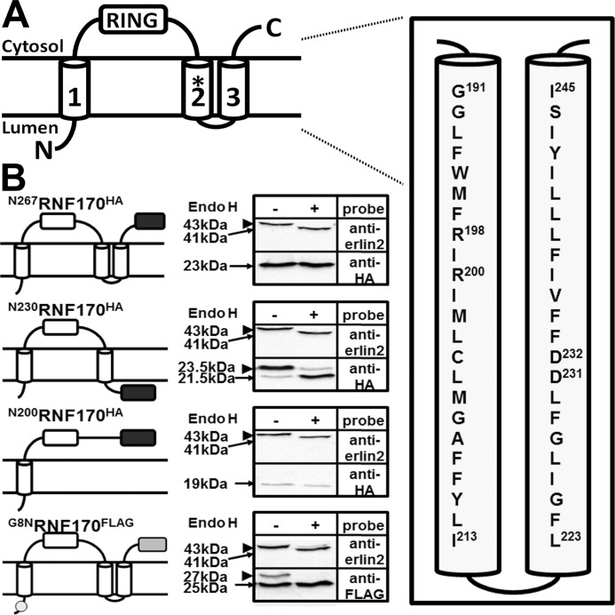
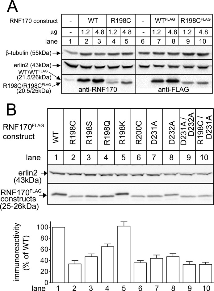
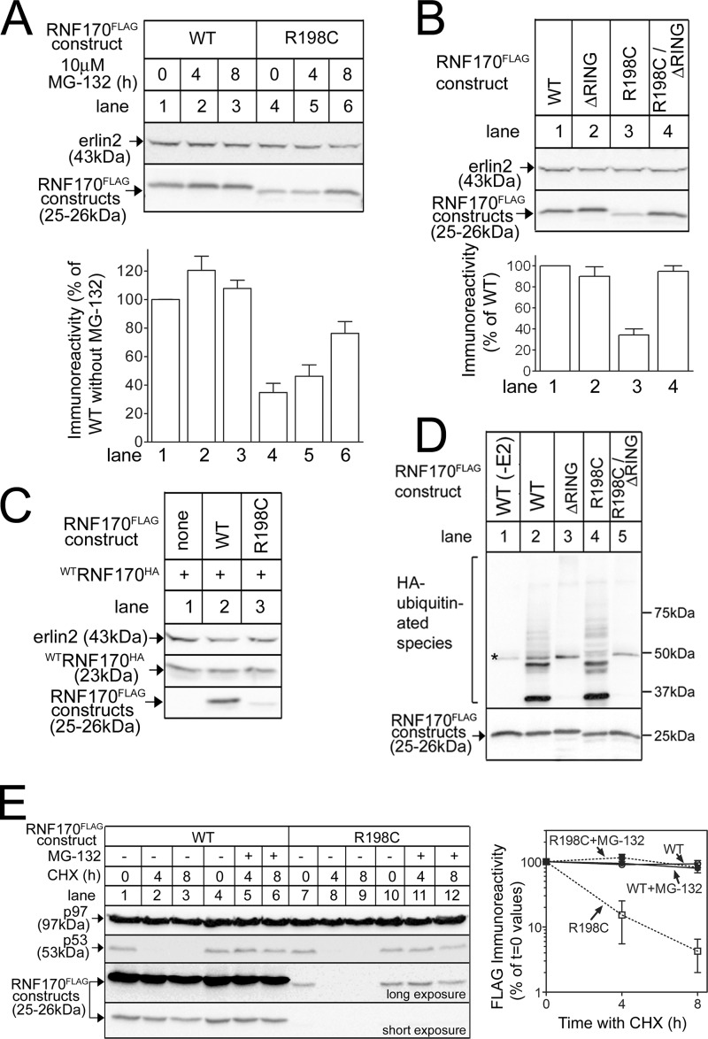
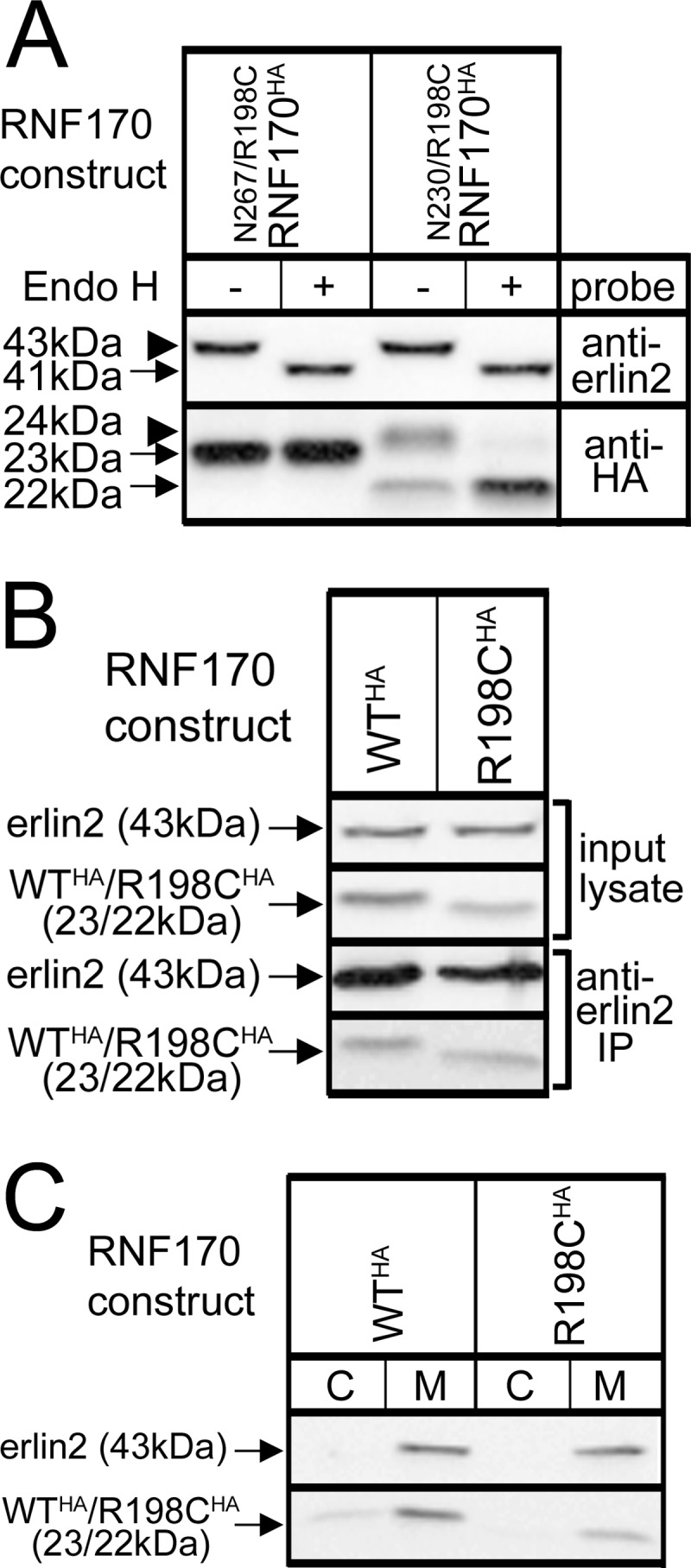
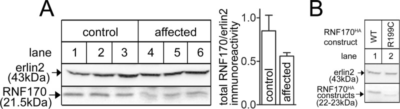
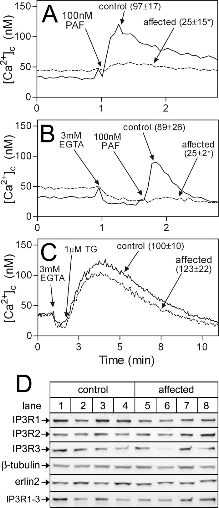
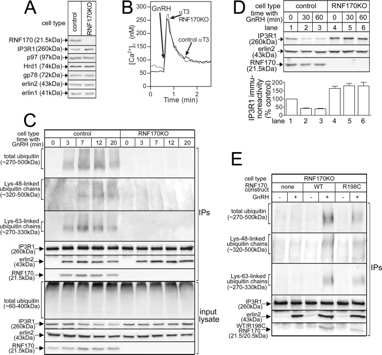
RNF170 is an endoplasmic reticulum membrane ubiquitin ligase that contributes to the ubiquitination of activated inositol 1,4,5-trisphosphate (IP3) receptors, and also, when point mutated (arginine to cysteine at position 199), causes autosomal dominant sensory ataxia (ADSA), a disease characterized by neurodegeneration in the posterior columns of the spinal cord. Here we demonstrate that this point mutation inhibits RNF170 expression and signaling via IP3 receptors. Inhibited expression of mutant RNF170 was seen in cells expressing exogenous RNF170 constructs and in ADSA lymphoblasts, and appears to result from enhanced RNF170 autoubiquitination and proteasomal degradation. The basis for these effects was probed via additional point mutations, revealing that ionic interactions between charged residues in the transmembrane domains of RNF170 are required for protein stability. In ADSA lymphoblasts, platelet-activating factor-induced Ca(2+) mobilization was significantly impaired, whereas neither Ca(2+) store content, IP3 receptor levels, nor IP3 production were altered, indicative of a functional defect at the IP3 receptor locus, which may be the cause of neurodegeneration. CRISPR/Cas9-mediated genetic deletion of RNF170 showed that RNF170 mediates the addition of all of the ubiquitin conjugates known to become attached to activated IP3 receptors (monoubiquitin and Lys(48)- and Lys(63)-linked ubiquitin chains), and that wild-type and mutant RNF170 have apparently identical ubiquitin ligase activities toward IP3 receptors. Thus, the Ca(2+) mobilization defect seen in ADSA lymphoblasts is apparently not due to aberrant IP3 receptor ubiquitination. Rather, the defect likely reflects abnormal ubiquitination of other substrates, or adaptation to the chronic reduction in RNF170 levels.

Keywords: E3 ubiquitin ligase; RNF170; calcium intracellular release; endoplasmic reticulum (ER); inositol trisphosphate receptor (InsP3R); neurodegenerative disease.


Figures

FIGURE 1.
FIGURE 1.
Membrane topology of RNF170. A, predicted topology of RNF170 (15) with the TM2/3 region expanded to show the mouse amino acid sequence. Note that the amino acid that corresponds to Arg199 of human RNF170 is Arg198 in mouse RNF170, and that the sequences of human and mouse TM2/3 regions are identical, with the exception that Met202 of mouse RNF170 is Ile203 in human RNF170 (15, 16). The RING domain, the three TM domain regions, and the N and C termini are indicated, and Arg198 is identified with an asterisk. The precise limits of the predicted TM domains have not been defined experimentally, but the scheme shown is predicted by multiple programs (e.g. TMHMM, TOPCONS, etc). B, N-glycosylation of RNF170 mutants. An HA/glycosylation tag (black box) was introduced at the C terminus of full-length RNF170 (N267RNF170HA), or truncated RNF170 lacking putative TM domain 3 (N230RNF170HA), or putative TM domains 2 and 3 (N200RNF170HA). These, and G8NRNF170FLAG (FLAG tag indicated by a gray box and the G8N mutation with a circle) were expressed in HeLa cells, lysates were incubated without or with 1 unit/μl of endo H for 3 h at 37 °C, and samples were subjected to SDS-PAGE. Blots were then probed with anti-HA or anti-FLAG to identify the exogenous RNF170 constructs, or anti-erlin2 to identify endogenous erlin2, which is known to be N-glycosylated (13, 14), and which serves as a positive control for endo H. The migration positions of unmodified and N-glycosylated species are indicated with arrows and arrowheads, respectively; degylcosylation of erlin2, N230RNF170HA, and G8NRNF170FLAG by endo H reduces their apparent molecular masses by ∼2 kDa.
FIGURE 2.
FIGURE 2.
Effects of mutation of Arg198 and other amino acids on RNF170 expression. cDNAs encoding wild-type (WT) and mutant RNF170 constructs and vector alone were transfected into HeLa cells and cell lysates were probed as indicated. Erlin2 and β-tubulin served as loading controls. A, lysates were probed with anti-RNF170, which detects both endogenous and exogenous untagged RNF170 constructs (lanes 1–5), or with anti-FLAG, which detects just exogenous FLAG-tagged constructs (lanes 6–10). B, lysates were probed with anti-FLAG and the histogram shows combined quantitated immunoreactivity (mean ± S.E., n ≥ 3).
FIGURE 3.
FIGURE 3.
The R198C mutation reduces RNF170 expression via autoubiquitination and the proteasome. A-C, cDNAs encoding tagged WT and mutant RNF170 constructs transfected into HeLa cells were treated as indicated, and cell lysates were probed with anti-FLAG or anti-HA to recognize exogenous RNF170 constructs, or anti-erlin2, which served a loading control. The histograms show combined quantitated immunoreactivity (mean ± S.E., n ≥ 4). D, RNF170FLAG constructs were immunopurified from transfected HeLa cells and incubated with E1 (UBE1), E2 (UbcH5b), and HA-ubiquitin as indicated for 30 min at 30 °C, with the exception of lane 1, which lacked E2. Samples were then probed with anti-HA to assess ubiquitination (upper panel), or anti-FLAG to assess the levels of RNF170FLAG constructs (lower panel). The asterisk marks a background band. E, transfected HeLa cells were treated as indicated with 20 μg/ml of cycloheximide (CHX), without or with 10 μm MG-132. Cell lysates were then probed with anti-p53 as a positive control for cycloheximide action, anti-p97 as a loading control, and anti-FLAG to recognize exogenous RNF170 constructs (long and short exposures are shown to facilitate visualization of immunoreactivity changes). The graph shows combined quantitated FLAG immunoreactivity, using the long exposure for R198CRNF170FLAG and the short exposure for WTRNF170FLAG (mean ± range, n = 2).
FIGURE 4.
FIGURE 4.
Lack of effect of the R198C mutation on RNF170 membrane association and topology, and interaction with the erlin1/2 complex. cDNAs encoding HA-tagged WT and mutant RNF170 constructs were transfected into HeLa cells. A, N-glycosylation of N267/R198CRNF170HA and N230/R198CRNF170HA, R198C-containing versions of the constructs shown in Fig. 1B, was assessed as described in the legend to Fig. 1B. B, interaction with the erlin1/2 complex. Erlin1/2 complex was immunoprecipitated with anti-erlin2 was probed for RNF170 constructs (lower panels). Note that the amounts of WTRNF170HA and R198CRNF170HA that co-immunoprecipitate are proportional to the amounts in input lysates, indicating that they interact with the erlin1/2 complex equally well. C, cells were lysed and centrifuged into cytosol (C) and membrane (M) factions as described (13), and probed as indicated.
FIGURE 5.
FIGURE 5.
RNF170 levels in lymphoblasts from control and ADSA-affected individuals. A, lysates from 3 control (lanes 1–3) and 3 affected individuals (lanes 4–6) were probed with anti-RNF170 and anti-erlin2. For the panels shown, immunoreactivity was quantitated and is plotted as total RNF170/erlin2 immunoreactivity (arbitrary units). Multiple quantitations of total RNF170 immunoreactivity from these and other lymphoblast lines showed that affected individuals contained 73 ± 5% of the immunoreactivity seen in control lymphoblasts (n ≥ 5). B, cDNAs encoding human WTRNF170HA and R199CRNF170HA were transfected into HeLa cells and cell lysates were probed as indicated. Erlin2 served as a loading control.
FIGURE 6.
FIGURE 6.
Assessment of the IP3-medited Ca2+ signaling pathway in lymphoblasts. Multiple control and affected lymphoblast cell lines (n) were analyzed. A–C, fura2-loaded cells were exposed to PAF, EGTA, and thapsigargin (TG) as indicated and [Ca2+]c was calculated. Values in parentheses are mean ± S.E. of PAF- or TG-induced increases in [Ca2+]c over basal values (n ≥ 4, with * indicating p < 0.05 when comparing values from control versus affected cells). D, lysates from 4 control and 4 affected lymphoblast lines were probed for the proteins indicated. Total IP3R immunoreactivity in control and affected lymphoblasts, measured with anti-IP3R1–3 (lowest panel), was 80 ± 20 and 76 ± 15 arbitrary units, respectively (mean ± S.E.).
FIGURE 7.
FIGURE 7.
CRISPR/Cas9-mediated deletion of RNF170 and reconstitution with exogenous RNF170 constructs. A, levels of RNF170, IP3R1, and other pertinent proteins in lysates from αT3 control and RNF170KO cells. B, GnRH (0.1 μm)-induced Ca2+ mobilization in αT3 control and RNF170KO cells. C, IP3R1 ubiquitination in αT3 control and RNF170KO cells. Cells were incubated with 0.1 μm GnRH and anti-IP3R1 IPs and input lysates were probed for the proteins indicated. D, IP3R1 down-regulation in αT3 control and RNF170KO cells. Cells were incubated with 0.1 μm GnRH and lysates were probed for the proteins indicated. The histogram shows combined quantitated immunoreactivity (mean ± S.E., n = 4). E, reconstitution of IP3R1 ubiquitination in RNF170KO cells. αT3 RNF170KO cells stably expressing WTRNF170 or R198CRNF170 were incubated without or with 0.1 μm GnRH for 20 min, and anti-IP3R1 IPs were probed for the proteins indicated.

References

    1. Finley D. (2009) Recognition and processing of ubiquitin-protein conjugates by the proteasome. Annu. Rev. Biochem. 78, 477–513 - PMC - PubMed
    1. Kleiger G., Mayor T. (2014) Perilous journey: a tour of the ubiquitin-proteasome system. Trends Cell Biol. 24, 352–359 - PMC - PubMed
    1. Deshaies R. J., Joazeiro C. A. (2009) RING Domain E3 ubiquitin ligases. Annu. Rev. Biochem. 78, 399–434 - PubMed
    1. Budhidarmo R., Nakatani Y., Day C. L. (2012) RINGs hold the key to ubiquitin transfer. Trends Biochem. Sci. 37, 58–65 - PubMed
    1. Metzger M. B., Pruneda J. N., Klevit R. E., Weissman A. M. (2014) RING-type E3 ligases: master manipulators of E2 ubiquitin-conjugating enzymes and ubiquitination. Biochim. Biophys. Acta 1843, 47–60 - PMC - PubMed

Publication types

MeSH terms

Supplementary concepts

LinkOut - more resources