. 2015 May 29;290(22):13958-71.
doi: 10.1074/jbc.M114.634535. Epub 2015 Apr 16.

CRL3IBTK Regulates the Tumor Suppressor Pdcd4 through Ubiquitylation Coupled to Proteasomal Degradation

Affiliations

CRL3IBTK Regulates the Tumor Suppressor Pdcd4 through Ubiquitylation Coupled to Proteasomal Degradation

Antonio Pisano et al. J Biol Chem. .

Abstract

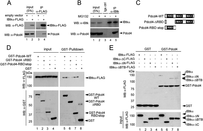
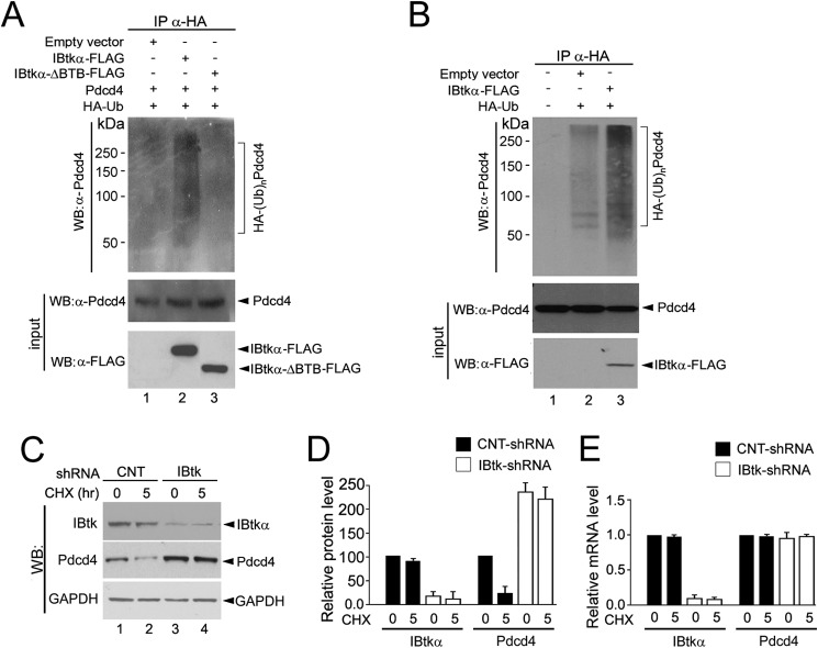
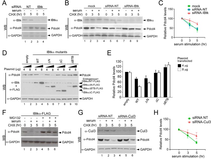
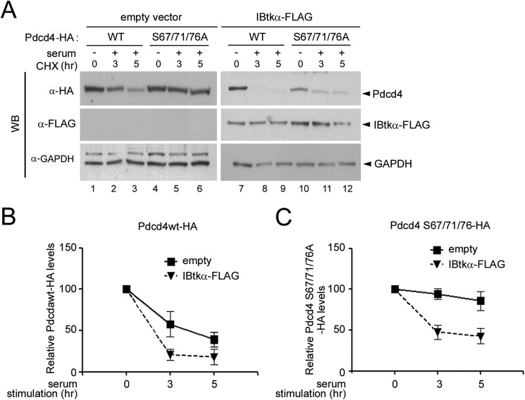
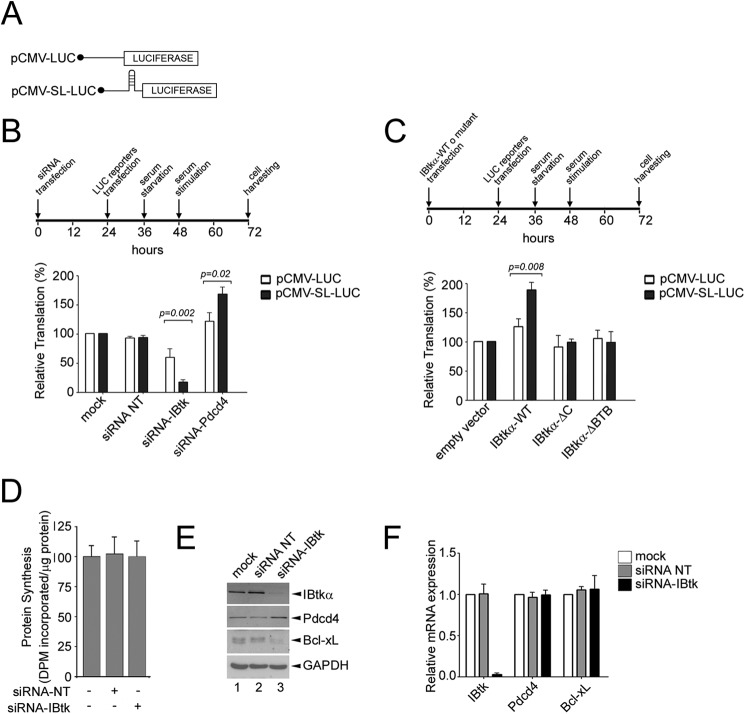
The human inhibitor of Bruton's tyrosine kinase isoform α (IBtkα) is a BTB protein encoded by the IBTK gene, which maps to chromosomal locus 6q14.1, a mutational hot spot in lymphoproliferative disorders. Here, we demonstrate that IBtkα forms a CRL3(IBTK) complex promoting its self-ubiquitylation. We identified the tumor suppressor Pdcd4 as IBtkα interactor and ubiquitylation substrate of CRL3(IBTK) for proteasomal degradation. Serum-induced degradation of Pdcd4 required both IBtkα and Cul3, indicating that CRL3(IBTK) regulated the Pdcd4 stability in serum signaling. By promoting Pdcd4 degradation, IBtkα counteracted the suppressive effect of Pdcd4 on translation of reporter luciferase mRNAs with stem-loop structured or unstructured 5'-UTR. IBtkα depletion by RNAi caused Pdcd4 accumulation and decreased the translation of Bcl-xL mRNA, a well known target of Pdcd4 repression. By characterizing CRL3(IBTK) as a novel ubiquitin ligase, this study provides new insights into regulatory mechanisms of cellular pathways, such as the Pdcd4-dependent translation of mRNAs.

Keywords: E3 ubiquitin ligase; IBtk; Pdcd4; serum; translation regulation; tumor suppressor gene; ubiquitylation (ubiquitination).


Figures

FIGURE 1.
FIGURE 1.
A, Clustal Omega program-based analysis of identity and similarity of human IBtkα and yeast Btb1 proteins. Yellow boxes highlight the BTB domains. *, identical positions, which have a single, fully conserved residue; colon and period, similar positions, which have conservation between groups of amino acids with strongly or weakly similar properties, respectively. B, schematic representation of the domain organization of human IBtkα and yeast Btb1 proteins (data from SwissProt, InterPro). C, superposition of the 3-box motifs of IBtkα and SPOP was elaborated by using Phyre2 software.
FIGURE 2.
FIGURE 2.
IBtkα assembles within a CRL3 ubiquitin ligase complex in vivo. A, association of ectopically expressed IBtkα and Cul3. HEK293T cells (3 × 10⁶) were transfected with IBtkα-FLAG (4 μg) or Myc-Cul3 (4 μg) or left untransfected. Forty-eight hours later, cell extracts (1 mg) were immunoprecipitated with the indicated antibodies and analyzed by WB. B, association of endogenous IBtkα and Cul3. HeLa cells (3 × 10⁶) were transfected with IBtkα-FLAG (4 μg) or left untransfected. The following steps were performed as indicated in A. C, schematic representation of IBtkα mutants used in this study. D, BTB domains of IBtkα are required for binding to Cul3. HEK293T cells (3 × 10⁶) were transfected with Myc-Cul3 (4 μg) and wild type or mutant IBtkα-FLAG (4 μg) or left untransfected. The following steps were performed as indicated in A. E, IBtkα binds to the amino terminus of Cul3. HEK293T cells (3 × 10⁶) were transfected with IBtkα-FLAG (4 μg), Myc-Cul3 (4 μg), or Myc-Cul3ΔN41 (4 μg) or left untransfected. The following steps were performed as indicated in A. *, nonspecific bands. F, IBtkα generates a macromolecular complex with Cul3 and Rbx1. HEK293T (3 × 10⁶) cells were transfected with IBtkα-FLAG (4 μg), Myc-Cul3 (4 μg), and HA-Rbx1 (4 μg). The following steps were performed as indicated in A.
FIGURE 3.
FIGURE 3.
IBtkα undergoes Cul3-dependent Lys48 polyubiquitylation and proteasomal degradation. A, Cul3 mediates the polyubiquitylation of IBtkα. HEK293T cells (3 × 10⁶) were transfected with His-tagged ubiquitin (His-Ub; 12 μg), with or without IBtkα-FLAG (4 μg), Myc-Cul3 (4 μg), or DN-hCul3-FLAG (4 μg) expression vectors. Forty-eight hours after transfection, cells were treated with MG132 (20 μm) for 4 h before lysis. Cell extracts were pulled down with cobalt-agarose affinity chromatography, resolved by 6% SDS-PAGE, and analyzed by WB with anti-FLAG antibody to detect IBtkα-FLAG ubiquitylated isoforms. B, IBtkα undergoes Lys48 polyubiquitylation in vivo. HEK293T cells (3 × 10⁶) were transfected with IBtkα-FLAG (4 μg) or the corresponding empty vector (4 μg), and 48 h later, cells were treated with MG132 (20 μm) for 4 h before lysis. Cell extracts were immunoprecipitated with anti-FLAG antibody and resolved by 6% SDS-PAGE, followed by immunoblotting with anti-Ub Lys48 or anti-Ub Lys63 antibody. C, evaluation of IBtkα half-life. HEK293T cells (3 × 10⁶) were treated for 1 h with MG132 (20 μm) or vehicle and then incubated with CHX (100 μg/ml) for up to 16 h. Cell lysates (30 μg) were separated by NuPAGE 4–12% SDS-PAGE and analyzed by WB with the indicated antibodies. D, quantification of IBtkα half-life. Protein band intensities of the experiment described in C were normalized to the corresponding GAPDH intensity and then normalized to the 0 h time point (100%). The mean densitometric values ± S.D. (error bars) of three independent experiments are shown.
FIGURE 4.
FIGURE 4.
IBtkα interacts with Pdcd4. A, in vivo association of IBtkα-FLAG with Pdcd4. HEK293T cells (3 × 10⁶) were transfected with IBtkα-FLAG or empty vector (4 μg). Forty-eight hours after transfection, cell lysates were immunoprecipitated with anti-FLAG antibody, and immunocomplexes were analyzed by WB with the indicated antibodies. B, in vivo association of endogenous IBtkα and Pdcd4 proteins. HEK293T cells (3 × 10⁶) were treated for 4 h with MG132 (20 μm) or vehicle. Cell lysates were immunoprecipitated with anti-IBtk or IgG antibodies, and immunocomplexes were analyzed by WB with the indicated antibodies. C, schematic representation of Pdcd4 wild type and mutants. D, mapping of Pdcd4 binding site for IBtkα. HEK293T cells (3 × 10⁶) were transfected with IBtkα-FLAG (4 μg), and cell lysates were subjected to GST pull-down, using GST-Pdcd4-WT, GST-Pdcd4DRBD, or GST-Pdcd4RBDStop recombinant proteins as baits. The pull-down was analyzed by WB with the indicated antibodies. E, mapping of IBtkα binding site for Pdcd4. HEK293T cells (3 × 10⁶) were transfected with IBtkα-FLAG or IBtkα-FLAG deletion mutants (4 μg). Cell lysates were subjected to GST-Pdcd4 pull-down and analyzed by WB with anti-FLAG antibody.
FIGURE 5.
FIGURE 5.
IBtkα promotes the Pdcd4 polyubiquitylation and degradation. A, IBtkα promotes the polyubiquitylation of exogenously expressed Pdcd4. HEK293T cells (3 × 10⁶) were transfected with Pdcd4 (4 μg) with or without IBtkα-FLAG, IBtkαΔBTB-FLAG, HA-tagged ubiquitin, or empty vector (4 μg). Forty-eight hours later, cells were treated with MG132 (20 μm) for 4 h before lysis. Cell extracts were subjected to IP with anti-HA antibody, and immunocomplexes were resolved by SDS-PAGE on a 6% gel, followed by WB with the indicated antibodies. B, IBtkα promotes the polyubiquitylation of endogenous Pdcd4 in vivo. C, IBtkα RNA interference increases the Pdcd4 protein content. HeLa cells (3 × 10⁶) were transduced with control shRNA (CNT-shRNA), or IBtk shRNA for 48 h and then incubated with CHX (100 μg/ml) for up to 5 h. Cell lysates (30 μg) were analyzed by WB with the indicated antibodies. D, densitometric analysis of WB protein bands relative to the experiment described in C. Optical density of WB protein bands was expressed as arbitrary units normalized to control shRNA taken as a value of 100. Mean values ± S.D. (error bars) of three independent experiments are shown. E, IBtkα RNA interference does not affect the Pdcd4 mRNA level. HeLa cells were transduced as described in C, total RNA was extracted 48 h later, and IBtkα and Pdcd4 transcripts were measured by quantitative RT-PCR. Relative mRNA levels were expressed as arbitrary units normalized to control shRNA taken as 1.0. Mean values ± S.D. of three independent experiments are shown.
FIGURE 6.
FIGURE 6.
IBtkα promotes the Pdcd4 proteasomal degradation upon serum stimulation. A, serum deprivation does not affect the IBtkα-dependent Pdcd4 degradation. HeLa cells (3 × 10⁶) were transfected with IBtk siRNA or control siRNA (siRNA-NT). Forty-eight hours later, cells were incubated with CHX (100 μg/ml) and serum-starved for up to 5 h. Cell lysates (30 μg) were separated by 4–12% NuPAGE and analyzed by WB with the indicated antibodies. B, IBtkα RNA interference counteracts the serum-induced Pdcd4 degradation in starved cells. HeLa cells (3 × 10⁶) were transfected with IBtk siRNA, control siRNA, or left untransfected (mock). Forty-eight hours later, cells were serum-starved for 16 h (time point 0) and then replenished with serum (10%) and incubated with CHX (100 μg/ml) for up to 5 h. The following steps were as described in A. C, quantification of Pdcd4 half-life with or without IBtkα RNA interference upon serum addition in starved cells. Protein band intensities of the experiment described in B were normalized to the corresponding GAPDH intensity and then compared with the 0 h time point. The mean densitometric values ± S.D. (error bars) of three independent experiments are shown. D, overexpression of IBtkα augments the degradation of Pdcd4. HeLa cells (3 × 10⁶) were transfected with two amounts (4 or 8 μg) of IBtkα-FLAG or deletion mutants or empty vector. Forty-eight hours later, cell lysates (30 μg) were separated by NuPAGE 4–12% and analyzed by WB with the indicated antibodies. E, quantification of the Pdcd4 level of the experiment described in D. Protein bands were normalized to the corresponding GAPDH intensity. The mean densitometric values ± S.D. of three independent experiments are shown. F, MG132 proteasome inhibitor rescues Pdcd4 from IBtkα-mediated degradation. HeLa cells (3 × 10⁶) were transfected with IBtkα-FLAG or empty vector (4 μg) and 48 h later were serum-starved for 16 h and then replenished with serum (10%) and incubated with MG132 (20 μm) for up to 5 h. G, Cul3 RNA interference counteracts the serum-induced Pdcd4 degradation. HeLa cells (3 × 10⁶) were transfected with Cul3 siRNA or control siRNA and subjected to serum starvation/addition and CHX treatment, as detailed in B. Cell extracts were analyzed by WB with the indicated antibodies. H, quantification of Pdcd4 half-life of the experiment described in G. Protein bands were normalized to the corresponding GAPDH intensity and then compared with the 0 h time point. The mean densitometric values ± S.D. of three independent experiments are shown.
FIGURE 7.
FIGURE 7.
IBtkα-mediated ubiquitylation of Pdcd4 does not depend on serines 71 and 76. A, overexpression of IBtkα induces the degradation of Pdcd4 S67A/S71A/S76A-HA mutant. HeLa cells (3 × 10⁶) cells were transfected with Pdcd4-WT-HA (2 μg) or Pdcd4 S67A/S71A/S76A-HA (2 μg) expression vectors, with or without IBtkα-FLAG (4 μg) or empty (4 μg) vector. Forty-eight hours later, cells were serum-starved for 16 h (time point 0) and then replenished with serum (10%) and incubated with CHX (100 μg/ml) for up to 5 h. Cell lysates (30 μg) were separated by NuPAGE 4–12% and analyzed by WB with the indicated antibodies. B and C, quantification of Pdcd4-WT-HA (B) or Pdcd4 S67A/S71A/S76A-HA (C) half-life with or without IBtkα overexpression upon serum addition in starved cells. Protein band intensities of the experiment described in A were normalized to the corresponding GAPDH intensity and then compared with the 0 h time point. The mean densitometric values ± S.D. (error bars) of three independent experiments are shown.
FIGURE 8.
FIGURE 8.
IBtkα enhances translation by counteracting the Pdcd4 repression of target mRNAs. A, schematic representation of luciferase reporter mRNAs. B, depletion of IBtkα by RNA interference decreases the translation of reporter mRNAs with stem-loop structured or unstructured 5′-UTR. HeLa cells (3 × 10⁶) cells were transfected with IBtk siRNA, Pdcd4 siRNA, or control siRNA or left untransfected (mock). After 24 h, cells were transfected with pCMV-LUC (0.2 μg) or pCMV-SL-LUC (0.2 μg) and serum-starved for 12 h, followed by growth in complete medium (10% FBS) for additional 24 h. The luciferase activity measured in untransfected cells was designated as 100%. Mean values ± S.D. (error bars) of five independent experiments are shown. C, overexpression of IBtkα enhances the translation of reporter mRNAs with stem-loop structured or unstructured 5′-UTR. HeLa cells (3 × 10⁶) were transfected with IBtkα-FLAG, IBtkα-FLAG mutants, or empty vector (4 μg). Subsequent steps were performed as described in B. D, IBtkα RNA interference does not affect the global protein synthesis. HeLa cells (3 × 10⁶) were transfected with IBtk siRNA or control siRNA or left untransfected (mock). The rate of protein synthesis was measured by incorporation of 35S-labeled methionine and cysteine into translated protein and normalized to total protein concentration. E, IBtkα depletion by RNA interference decreases the intracellular amount of Bcl-xL. HeLa cells (3 × 10⁶) were transfected with IBtk siRNA or control siRNA or left untransfected (mock), and 48 h later, cell lysates were analyzed by WB with the indicated antibodies. F, HeLa cells were transfected as described in E, and total RNA was analyzed by real-time PCR to measure the level of the indicated transcripts. Mean values ± S.D. of three independent experiments are shown.

References

    1. Hershko A., Ciechanover A. (1998) The ubiquitin system. Annu. Rev. Biochem. 67, 425–479 - PubMed
    1. Pickart C. M. (2004) Back to the future with ubiquitin. Cell 116, 181–190 - PubMed
    1. Lipkowitz S., Weissman A. M. (2011) RINGs of good and evil: RING finger ubiquitin ligases at the crossroads of tumour suppression and oncogenesis. Nat. Rev. Cancer 11, 629–643 - PMC - PubMed
    1. Petroski M. D., Deshaies R. J. (2005) Function and regulation of cullin-RING ubiquitin ligases. Nat. Rev. Mol. Cell Biol. 6, 9–20 - PubMed
    1. Saha A., Deshaies R. J. (2008) Multimodal activation of the ubiquitin ligase SCF by Nedd8 conjugation. Mol. Cell 32, 21–31 - PMC - PubMed

Publication types

MeSH terms