. 2015 May 29;290(22):13935-47.
doi: 10.1074/jbc.M114.617431. Epub 2015 Apr 16.

Functional Effect of Pim1 Depends upon Intracellular Localization in Human Cardiac Progenitor Cells

Affiliations

Functional Effect of Pim1 Depends upon Intracellular Localization in Human Cardiac Progenitor Cells

Kaitlen Samse et al. J Biol Chem. .

Abstract

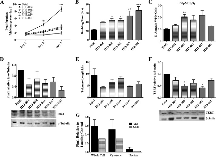
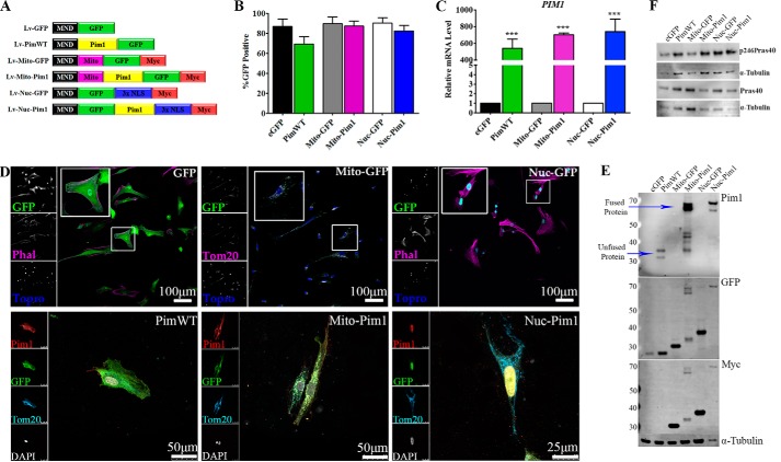
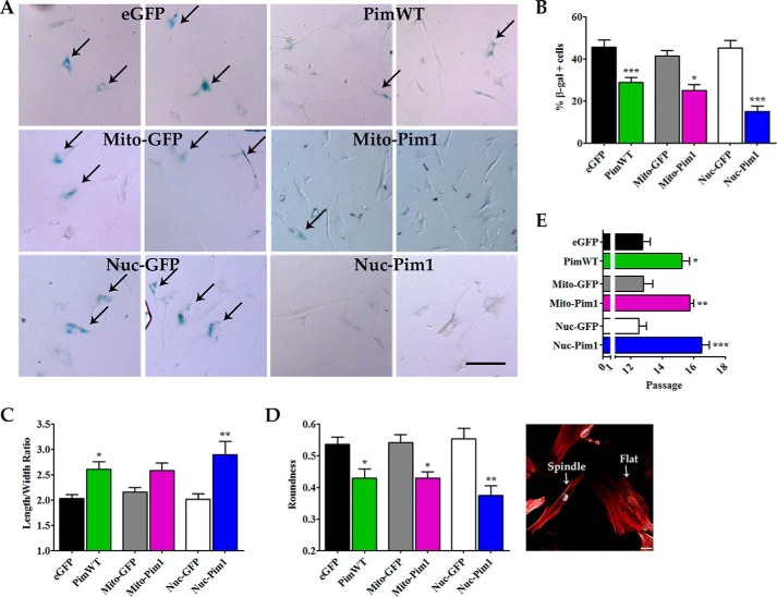
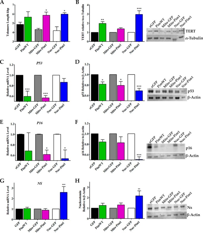
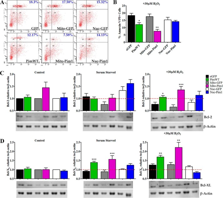
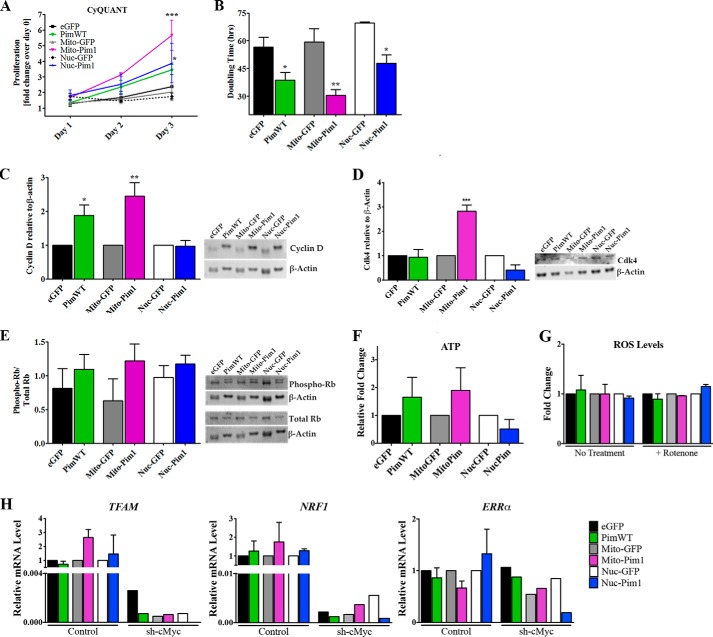
Human cardiac progenitor cells (hCPC) improve heart function after autologous transfer in heart failure patients. Regenerative potential of hCPCs is severely limited with age, requiring genetic modification to enhance therapeutic potential. A legacy of work from our laboratory with Pim1 kinase reveals effects on proliferation, survival, metabolism, and rejuvenation of hCPCs in vitro and in vivo. We demonstrate that subcellular targeting of Pim1 bolsters the distinct cardioprotective effects of this kinase in hCPCs to increase proliferation and survival, and antagonize cellular senescence. Adult hCPCs isolated from patients undergoing left ventricular assist device implantation were engineered to overexpress Pim1 throughout the cell (PimWT) or targeted to either mitochondrial (Mito-Pim1) or nuclear (Nuc-Pim1) compartments. Nuc-Pim1 enhances stem cell youthfulness associated with decreased senescence-associated β-galactosidase activity, preserved telomere length, reduced expression of p16 and p53, and up-regulation of nucleostemin relative to PimWT hCPCs. Alternately, Mito-Pim1 enhances survival by increasing expression of Bcl-2 and Bcl-XL and decreasing cell death after H2O2 treatment, thereby preserving mitochondrial integrity superior to PimWT. Mito-Pim1 increases the proliferation rate by up-regulation of cell cycle modulators Cyclin D, CDK4, and phospho-Rb. Optimal stem cell traits such as proliferation, survival, and increased youthful properties of aged hCPCs are enhanced after targeted Pim1 localization to mitochondrial or nuclear compartments. Targeted Pim1 overexpression in hCPCs allows for selection of the desired phenotypic properties to overcome patient variability and improve specific stem cell characteristics.

Keywords: Pim1; aging; apoptosis; heart failure; human cardiac progenitor cell; proliferation; senescence.


Figures

FIGURE 1.
FIGURE 1.
Characterization of hCPC. A, proliferation rate shows increases in fetal hCPCs versus adult hCPCs on days 2 and 3 as measured by CyQUANT assay. Bars refer to fast-growing fetal hCPCs (black), medium-growth rate adult hCPCs (black stripes), and slow-growing adult hCPCs (gray). B, population doubling time varies in different hCPC lines as measured by CyQUANT assay readings. C, adult hCPC are more susceptible to apoptosis than fetal CPC when treated with 30 μm H2O2 for 3 h as measured by FACS analysis of annexin V and PI staining. D, disparity in endogenous Pim1 expression of adult hCPC represented as fold-change over fetal CPC measured by immunoblot analysis (each sample was normalized to α-tubulin). E, telomere lengths decrease in hCPC as measured by qRT-PCR. F, TERT protein expression measured by immunoblot analysis shows variability in hCPC lines. G, endogenous Pim1 localization, as shown in whole cell versus cytosolic and nuclear subcompartments in fetal versus adult hCPCs, is shown by immunoblot analysis (whole cell and cytosolic fractions were normalized to α-tubulin and nuclear fractions were normalized to Lamin A/C). *, p < 0.05; **, p < 0.01; ***, p < 0.001. Significance is calculated for fetal versus adult hCPC.
FIGURE 2.
FIGURE 2.
hCPC engineered with Pim1. A, vectors driving expression of GFP (Lv-eGFP), GFP and Pim1 (Lv-PimWT), mitochondrial targeted GFP (Lv-Mito-GFP), mitochondrial targeted GFP and Pim1 (Lv-Mito-Pim1), nuclear-targeted GFP (Lv-Nuc-GFP), and nuclear-targeted GFP and Pim1 (Lv-Nuc-Pim1) were used in this study. B, percentage of GFP-positive hCPCs after lentiviral modification as measured by confocal microscopy. C, increased PIM1 gene expression in Pim1-modified hCPCs as measured by qPCR analysis. All samples normalized to 18S transcription. D, genetic modification confirmed by localization of GFP and/or Pim1 protein expression in engineered hCPCs by immunocytochemistry. E, expression of Pim1 and GFP confirmed by immunoblot analysis. F, kinase activity of Pim1 lentiviral constructs is intact, as shown by phosphorylation of PraS40 Thr-246 in engineered hCPCs. ***, p < 0.001.
FIGURE 3.
FIGURE 3.
Replicative senescence is antagonized by Nuc-Pim1. A, SA β-gal staining of engineered hCPC at passage 13. Scale bar = 100 μm. B, graphical representation of percent of SA β-gal positive cells at passage 13. C, Pim1 modification shows decreased roundness and increased length to width ratio of hCPC at passage 13. Image displays characteristic round, flat cell (right) and a long, spindle shaped cell (left). D, passage at which growth arrest occurs in hCPC, indicative of replicative senescence. *, p < 0.05; **, p < 0.01; ***, p < 0.001.
FIGURE 4.
FIGURE 4.
Aged adult hCPC are reverted to youthful phenotypes with nuclear targeted Pim1. A, telomere length measured by RT-qPCR indicates telomere elongation with Pim1 modification. B, increased protein expression of TERT is observed by immunoblot analysis in Nuc-Pim1 hCPC (each sample is normalized to α-Tubulin). C, E, and G, Nuc-Pim1 modification decreases mRNA levels of senescence-associated markers P53 and P16 and increases NS as measured by real time PCR (samples normalized to 18S). D, F, and H, immunoblot analysis for p53, p16, and Ns with graphical representation of fold-change as compared with eGFP, Mito-GFP, and Nuc-GFP (each sample is normalized to β-actin). *, p < 0.05; **, p < 0.01; ***, p < 0.001.
FIGURE 5.
FIGURE 5.
Mitochondrial targeted Pim1 enhances survival. A and B, percent annexin V/PI double positive cells after apoptotic stimuli of 30 μm H2O2 for 3 h as measured by FACS analysis. C, Bcl-2 protein expression in untreated control, serum starved and H2O2-treated hCPC by immunoblot analysis. D, protein expression of untreated control, serum starved, and H2O2-treated hCPC by immunoblot analysis. Graph analyses are of fold-change over eGFP, Mito-GFP, or Nuc-GFP untreated control for Bcl-2 and Bcl-XL expression (each sample normalized to β-actin). *, p < 0.05; **, p < 0.01; ***, p < 0.001.
FIGURE 6.
FIGURE 6.
Growth kinetics are increased by Mito-Pim1. A, increased proliferation rate as measured by CyQUANT analysis after Pim1 modification. B, doubling time decreases, as calculated by CyQUANT measurements, with Pim1 overexpression. C, protein expression of cell cycle regulator Cyclin D as measured by immunoblot analysis. Fold-change as relative to eGFP, Mito-GFP, or Nuc-GFP. D, immunoblot analysis depicts an increase in protein expression of Cdk4 in Mito-Pim1 hCPC relative to controls. E, graphical representation of immunoblot analysis shows fold-change of phospho-Rb relative to total Rb protein expression. Each sample was normalized to β-actin. F, increased ATP levels in PimWT and Mito-Pim1 hCPCs as meansured by a ATP bioluminescence assay. G, difference in ROS levels not apparent with Pim1 modification. H, increase in mitochondrial biogenesis genes is dependent upon cMyc. *, p < 0.05; **, p < 0.01; and ***, p < 0.001.

References

    1. Reinecke H., Minami E., Zhu W. Z., Laflamme M. A. (2008) Cardiogenic differentiation and transdifferentiation of progenitor cells. Circ. Res. 103, 1058–1071 - PMC - PubMed
    1. Fransioli J., Bailey B., Gude N. A., Cottage C. T., Muraski J. A., Emmanuel G., Wu W., Alvarez R., Rubio M., Ottolenghi S., Schaefer E., Sussman M. A. (2008) Evolution of the c-kit-positive cell response to pathological challenge in the myocardium. Stem Cells 26, 1315–1324 - PMC - PubMed
    1. Ellison G. M., Torella D., Dellegrottaglie S., Perez-Martinez C., Perez de Prado A., Vicinanza C., Purushothaman S., Galuppo V., Iaconetti C., Waring C. D., Smith A., Torella M., Cuellas Ramon C., Gonzalo-Orden J. M., Agosti V., Indolfi C., Galiñanes M., Fernandez-Vazquez F., Nadal-Ginard B. (2011) Endogenous cardiac stem cell activation by insulin-like growth factor-1/hepatocyte growth factor intracoronary injection fosters survival and regeneration of the infarcted pig heart. J. Am. Coll. Cardiol. 58, 977–986 - PubMed
    1. Beltrami A. P., Barlucchi L., Torella D., Baker M., Limana F., Chimenti S., Kasahara H., Rota M., Musso E., Urbanek K., Leri A., Kajstura J., Nadal-Ginard B., Anversa P. (2003) Adult cardiac stem cells are multipotent and support myocardial regeneration. Cell 114, 763–776 - PubMed
    1. Urbanek K., Torella D., Sheikh F., De Angelis A., Nurzynska D., Silvestri F., Beltrami C. A., Bussani R., Beltrami A. P., Quaini F., Bolli R., Leri A., Kajstura J., Anversa P. (2005) Myocardial regeneration by activation of multipotent cardiac stem cells in ischemic heart failure. Proc. Natl. Acad. Sci. U.S.A. 102, 8692–8697 - PMC - PubMed

Publication types

MeSH terms