. 2015 Apr 7;19(1):149.
doi: 10.1186/s13054-015-0883-z.

Human severe sepsis cytokine mixture increases β2-integrin-dependent polymorphonuclear leukocyte adhesion to cerebral microvascular endothelial cells in vitro

Affiliations

Human severe sepsis cytokine mixture increases β2-integrin-dependent polymorphonuclear leukocyte adhesion to cerebral microvascular endothelial cells in vitro

Chris Blom et al. Crit Care. .

Abstract

Introduction: Sepsis-associated encephalopathy (SAE) is a state of acute brain dysfunction in response to a systemic infection. We propose that systemic inflammation during sepsis causes increased adhesion of leukocytes to the brain microvasculature, resulting in blood-brain barrier dysfunction. Thus, our objectives were to measure inflammatory analytes in plasma of severe sepsis patients to create an experimental cytokine mixture (CM), and to use this CM to investigate the activation and interactions of polymorphonuclear leukocytes (PMN) and human cerebrovascular endothelial cells (hCMEC/D3) in vitro.

Methods: The concentrations of 41 inflammatory analytes were quantified in plasma obtained from 20 severe sepsis patients and 20 age- and sex-matched healthy controls employing an antibody microarray. Two CMs were prepared to mimic severe sepsis (SSCM) and control (CCM), and these CMs were then used for PMN and hCMEC/D3 stimulation in vitro. PMN adhesion to hCMEC/D3 was assessed under conditions of flow (shear stress 0.7 dyn/cm(2)).

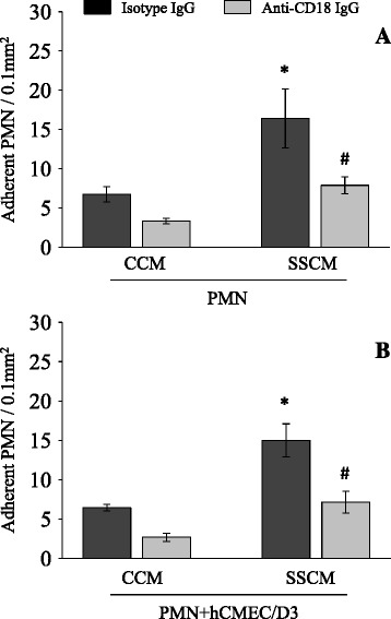
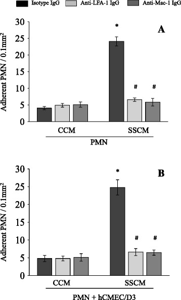
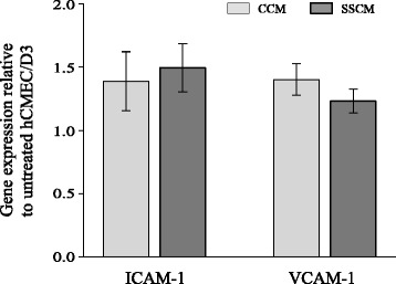
Results: Eight inflammatory analytes elevated in plasma obtained from severe sepsis patients were used to prepare SSCM and CCM. Stimulation of PMN with SSCM led to a marked increase in PMN adhesion to hCMEC/D3, as compared to CCM. PMN adhesion was abolished with neutralizing antibodies to either β2 (CD18), αL/β2 (CD11α/CD18; LFA-1) or αM/β2 (CD11β/CD18; Mac-1) integrins. In addition, immune-neutralization of the endothelial (hCMEC/D3) cell adhesion molecule, ICAM-1 (CD54) also suppressed PMN adhesion.

Conclusions: Human SSCM up-regulates PMN pro-adhesive phenotype and promotes PMN adhesion to cerebrovascular endothelial cells through a β2-integrin-ICAM-1-dependent mechanism. PMN adhesion to the brain microvasculature may contribute to SAE.


Figures

Figure 1
Figure 1
PMN adhesion to hCMEC/D3 following stimulation with severe sepsis cytokine mixture (SSCM). hCMEC/D3 were grown on laminar flow microchannels and interacted with PMN following stimulation of hCMEC/D3, PMN, or both PMN and hCMEC/D3, with CCM or SSCM. In these experiments, hCMEC/D3 were stimulated with CCM or SSCM for five hours. PMN were stimulated for 10 minutes immediately before interacting them with hCMEC/D3. PMN adhesion to hCMEC/D3 was assessed in the presence of flow (laminar shear stress 0.7 dyn/cm2). n = 7; *P <0.05 as compared to corresponding control cytokine mixture (CCM). CCM-control cytokine mixture; SSCM-severe sepsis cytokine mixture; hCMEC/D3-human cerebral microvascular endothelial cells/D3; PMN-polymorphonuclear leukocytes.
Figure 2
Figure 2
Effects of anti-β 2 -integrin (CD18) antibody on severe sepsis cytokine mixture (SSCM)-induced PMN adhesion to hCMEC/D3. PMN were first stimulated with CCM or SSCM for 10 minutes and subsequently treated with anti-β2-integrin (CD18) function neutralizing antibody (or control isotype antibody) for an additional 15 minutes. PMN adhesion to stimulated PMN (A) or stimulated PMN and hCMEC/D3 (B) was assessed in the presence of flow (laminar shear stress 0.7 dyn/cm2). n = 4. *P <0.05 as compared to corresponding control cytokine mixture (CCM); # P <0.05 as compared to isotype IgG treatment. CCM-control cytokine mixture; SSCM-severe sepsis cytokine mixture; hCMEC/D3-human cerebral microvascular endothelial cells/D3; PMN-polymorphonuclear leukocytes; IgG-immunoglobulin G; CD18-cluster of differentiation 18.
Figure 3
Figure 3
Effects of anti-LFA-1 and anti-Mac-1 antibodies on severe sepsis cytokine mixture (SSCM)-induced PMN adhesion to hCMEC/D3. PMN were first stimulated with CCM or SSCM for 10 minutes and subsequently treated with anti-LFA-1 or anti-Mac-1 function neutralizing antibody (or control isotype antibody) for an additional 15 minutes. PMN adhesion to stimulated PMN (A) or stimulated PMN and hCMEC/D3 (B) was assessed in the presence of flow (laminar shear stress 0.7 dyn/cm2). n = 4. *P <0.05 as compared to corresponding control cytokine mixture (CCM); # P <0.05 as compared to isotype IgG treatment. CCM-control cytokine mixture; SSCM-severe sepsis cytokine mixture; hCMEC/D3-human cerebral microvascular endothelial cells/D3; PMN-polymorphonuclear leukocytes; IgG-immunoglobulin G; LFA-1 - lymphocyte function-associated antigen 1; Mac-1 - macrophage-1 antigen.
Figure 4
Figure 4
Effects of anti-ICAM-1 antibody on severe sepsis cytokine mixture (SSCM)-induced PMN adhesion to hCMEC/D3. hCMEC/D3 were stimulated with CCM or SSCM for 5 hours, treated with anti-ICAM-1 function neutralizing antibody for 30 minutes, and interacted with CCM- or SSCM-stimulated (10 minutes) PMN to assess PMN adhesion in the presence of flow (laminar shear stress 0.7 dyn/cm2). n = 6; *P <0.05 as compared to corresponding control cytokine mixture (CCM); # P <0.05 as compared to isotype IgG treatment. CCM-control cytokine mixture; SSCM-severe sepsis cytokine mixture; hCMEC/D3-human cerebral microvascular endothelial cells/D3; PMN-polymorphonuclear leukocytes; Ab-antibody; ICAM-1 – intercellular adhesion molecule-1.
Figure 5
Figure 5
Effects of severe sepsis cytokine mixture (SSCM) on adhesion molecule ICAM-1 and VCAM-1 expression in hCMEC/D3. hCMEC/D3 were stimulated for 4 hours with either CCM or SSCM preceding analysis of VCAM-1 and ICAM-1 gene expression using qPCR. Data is presented as gene expression normalized to GAPDH and 18S expression, relative to hCMEC/D3 treated with serum-free VascuLife EnGS-Mv cell culture media (n = 5). CCM-control cytokine mixture; SSCM-severe sepsis cytokine mixture; hCMEC/D3-human cerebral microvascular endothelial cells/D3; ICAM-1 – intercellular adhesion molecule-1; VCAM-1 – vascular cell adhesion molecule-1; 18S-18S ribosomal RNA; GAPDH-glyceraldehyde 3-phosphate dehydrogenase; qPCR- quantitative polymerase chain reaction.

References

    1. Angus DC, Linde-Zwirble WT, Lidicker J, Clermont G, Carcillo J, Pinsky MR. Epidemiology of severe sepsis in the United States: analysis of incidence, outcome, and associated costs of care. Crit Care Med. 2001;29:1303–1310. doi: 10.1097/00003246-200107000-00002. - DOI - PubMed
    1. Bone RC, Balk RA, Cerra FB, Dellinger RP, Fein AM, Knaus WA, et al. Definitions for sepsis and organ failure and guidelines for the use of innovative therapies in sepsis. The ACCP/SCCM Consensus Conference Committee. American College of Chest Physicians/Society of Critical Care Medicine. Chest. 1992;101:1644–1655. doi: 10.1378/chest.101.6.1644. - DOI - PubMed
    1. Young GB, Bolton CF, Austin TW, Archibald YM, Gonder J, Wells GA. The encephalopathy associated with septic illness. Clin Invest Med. 1990;13:297–304. - PubMed
    1. Sprung CL, Peduzzi PN, Shatney CH, Schein RM, Wilson MF, Sheagren JN, et al. Impact of encephalopathy on mortality in the sepsis syndrome, The Veterans Administration Systemic Sepsis Cooperative Study Group. Crit Care Med. 1990;18:801–806. doi: 10.1097/00003246-199008000-00001. - DOI - PubMed
    1. Iwashyna TJ, Ely EW, Smith DM, Langa KM. Long-term cognitive impairment and functional disability among survivors of severe sepsis. JAMA. 2010;304:1787–1794. doi: 10.1001/jama.2010.1553. - DOI - PMC - PubMed

Publication types

MeSH terms