. 2015 Jul;5(7):740-51.
doi: 10.1158/2159-8290.CD-14-1347. Epub 2015 Apr 16.

In Vivo Role of INPP4B in Tumor and Metastasis Suppression through Regulation of PI3K-AKT Signaling at Endosomes

Affiliations

In Vivo Role of INPP4B in Tumor and Metastasis Suppression through Regulation of PI3K-AKT Signaling at Endosomes

Chen Li Chew et al. Cancer Discov. 2015 Jul.

Abstract

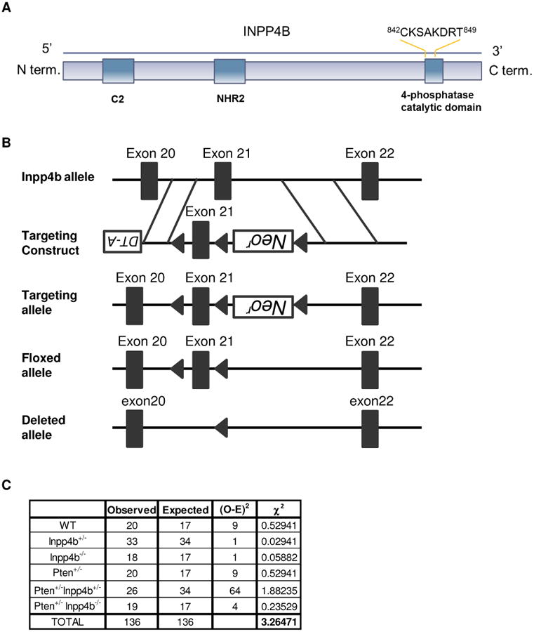
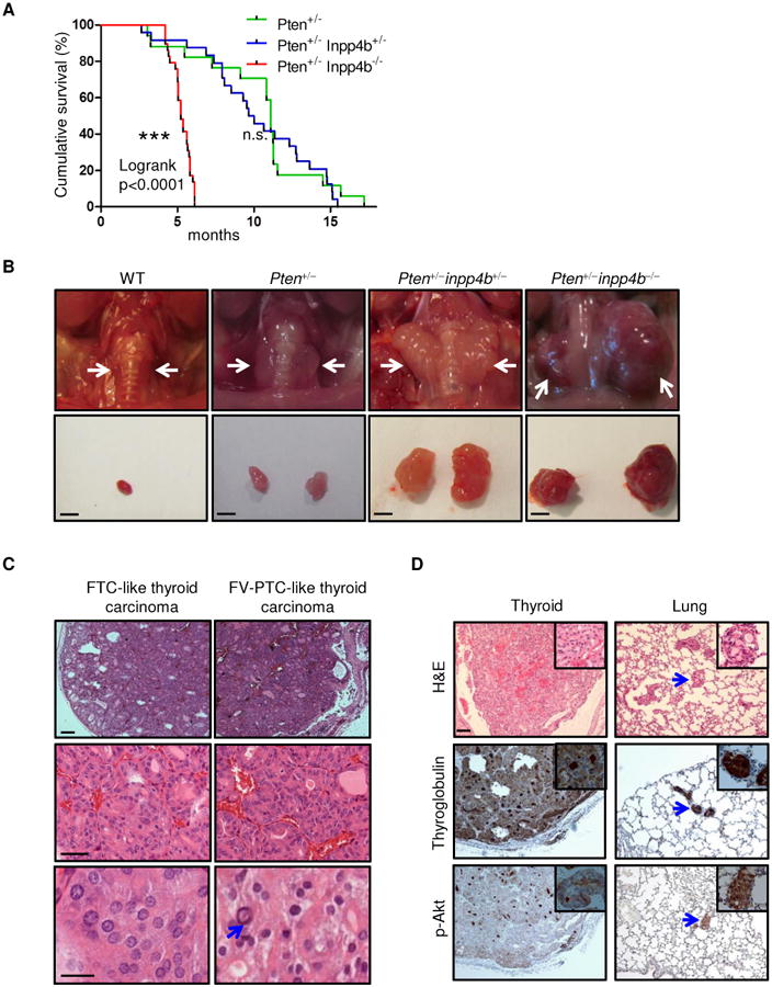
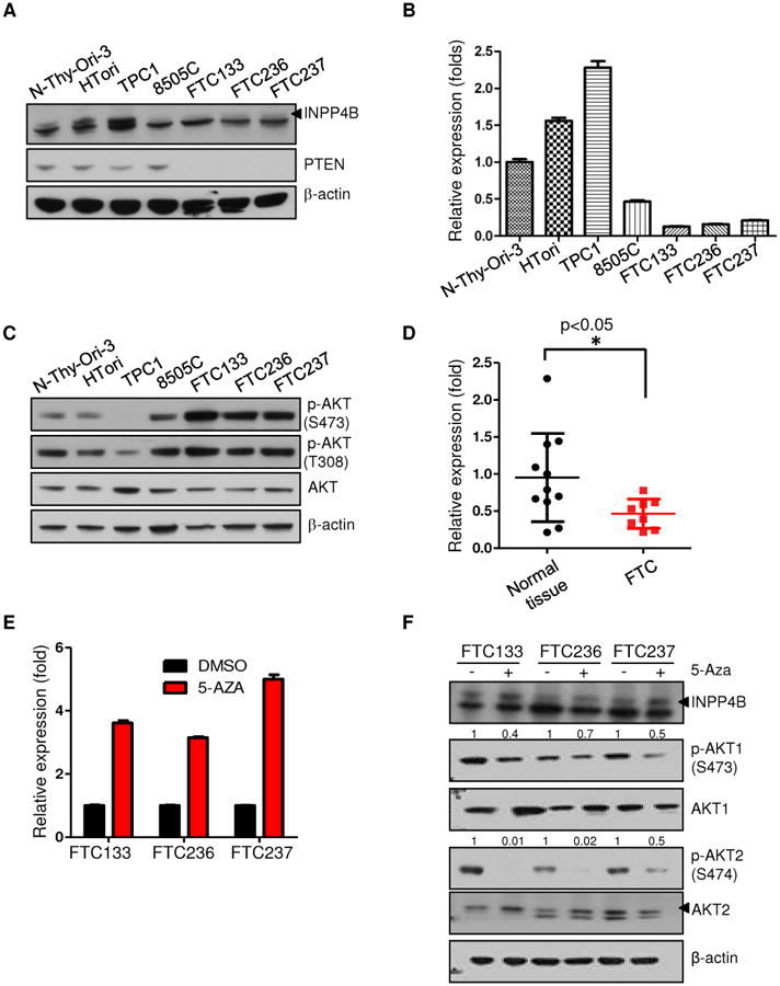
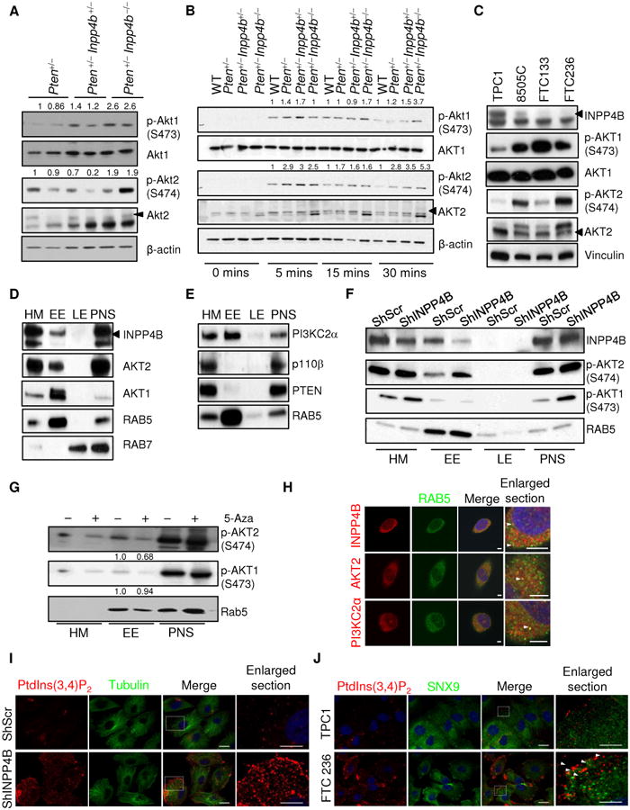
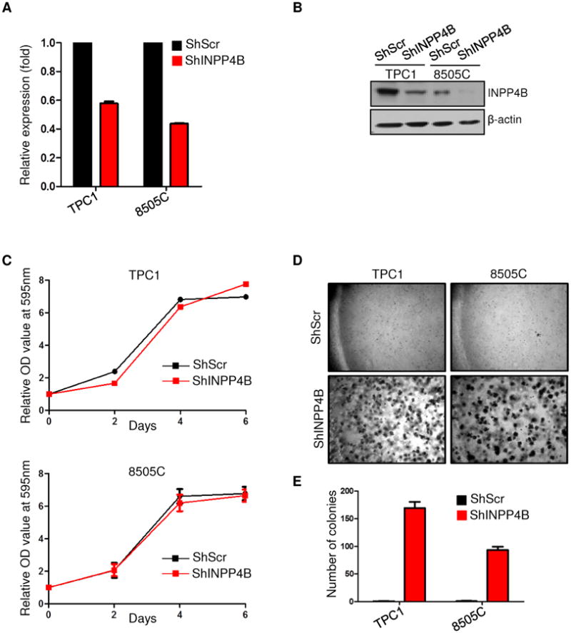
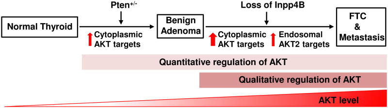
The phosphatases PTEN and INPP4B have been proposed to act as tumor suppressors by antagonizing PI3K-AKT signaling and are frequently dysregulated in human cancer. Although PTEN has been extensively studied, little is known about the underlying mechanisms by which INPP4B exerts its tumor-suppressive function and its role in tumorigenesis in vivo. Here, we show that a partial or complete loss of Inpp4b morphs benign thyroid adenoma lesions in Pten heterozygous mice into lethal and metastatic follicular-like thyroid cancer (FTC). Importantly, analyses of human thyroid cancer cell lines and specimens reveal INPP4B downregulation in FTC. Mechanistically, we find that INPP4B, but not PTEN, is enriched in the early endosomes of thyroid cancer cells, where it selectively inhibits AKT2 activation and in turn tumor proliferation and anchorage-independent growth. We therefore identify INPP4B as a novel tumor suppressor in FTC oncogenesis and metastasis through localized regulation of the PI3K-AKT pathway at the endosomes.

Significance: Although both PTEN and INPP4B can inhibit PI3K-AKT signaling through their lipid phosphatase activities, here we demonstrate lack of an epistatic relationship between the two tumor suppressors. Instead, the qualitative regulation of PI3K-AKT2 signaling by INPP4B provides a mechanism for their cooperation in suppressing thyroid tumorigenesis and metastasis.


Conflict of interest statement

Conflict of Interest: None of the authors have anything to declare

Figures

Figure 1
Figure 1. Generation and Characterization of Pten+/−Inpp4b+/− and Pten+/−Inpp4b−/− mice
A. Diagram representing the structure of INPP4B, which contains an N-terminus C2 domain and a C-terminus phosphatase domain harboring the phosphatase catalytic motif CX5R. B. Schematic map of the WT Inpp4b locus (top), targeting vector (upper middle) and predicted targeted allele (lower middle) and knockout allele (bottom). C. Table depicting the observed versus the expected numbers of mice of the respective genotypes from a Pten+/−Inpp4b+/− and Inpp4b+/− cross. These values gave a χ2 of 3.26, which is lower than the critical value of 11.070 which would yield an α=0.05. Thus, we conclude that the mutants were born following Mendelian frequencies.
Figure 2
Figure 2. Loss of Inpp4b in Pten+/− mice leads to follicular thyroid carcinoma
A. Kaplan-Meier survival curve of Pten+/−, Pten+/−Inpp4b+/− and Pten+/−Inpp4b−/− mice. B. Top panel: gross anatomy of representative thyroids from mice of the indicated genotypes. White arrows point to the location of the thyroid. Bottom panel: dissected thyroids from mice of the indicated genotypes; Scale bar, 2mm. C. Left panel: H&E staining of a follicular thyroid carcinoma (FTC)-like thyroid tumor which is thinly encapsulated with microfollicular architecture. Right panel: H&E staining of a follicular variant of papillary thyroid carcinoma (FV-PTC)-like thyroid tumor. Middle and bottom panels highlight nuclear atypia characteristic of FV-PTC, including intranuclear pseudoinclusions (arrow). Scale bars, top and middle panels: 100μm, bottom panel: 20 μm, D. Top panel: H&E staining of thyroid tumor and lung metastases from Pten+/−Inpp4b−/− mice; middle & bottom panel: thyroglobulin and p-Akt (Ser473) staining. Scale bar, 100μm. Insets show thyroid cancer cells. Blue arrows point to the location of the metastases in the lungs.
Figure 3
Figure 3. INPP4B expression is reduced in human follicular thyroid cell lines and human surgical specimens
A. Western blot analysis of thyroid cancer cell lines for INPP4B and PTEN expression. Arrow indicates specific band. The arrowhead indicates the specific band of INPP4B protein. B. RT-qPCR analysis of thyroid cancer cell lines for INPP4B transcript levels. C. Western blot analysis of thyroid cancer lines for AKT activation on both Serine 473 and Threonine 308 residues. D. RT-qPCR analysis of INPP4B in unmatched normal versus FTC patient tumor samples. E-F. Thyroid cancer cell lines were treated with 3uM 5-Aza-2′-deoxycytidine for 5 days. Panel shows transcript (E) and protein analysis of INPP4B expression and AKT activation levels in DMSO versus 5-Aza treated FTC cells. (F). Arrows indicate specific band (see also Methods). AKT2 antibody used is (5239).
Figure 4
Figure 4. Loss of Inpp4b in Pten+/− cells leads to increased Akt activation
A-B. Western blot analysis of lysates from Pten+/− (n=3; ∼10-11months), Pten+/−Inpp4b+/− (n=2; ∼6-8months) and Pten+/−Inpp4b−/− (n=3; ∼6 months) thyroid tissues (A), lysates from immortalized MEFs after serum starvation-restimulation. The MEFs were stimulated with serum for the indicated amount of time. (B). C. Western blot analysis of total lysate from different thyroid cancer cells. Please note that the AKT2 antibody used in A-B (5239) is different from C (3063), resulting in the appearance of different unspecific bands. Arrows indicate specific band (see also Methods). D-E. Western blot analysis of cell fractions from TPC1 cells. HM: cytosolic/heavy membranes; EE: early endosome: LE: late endosome; PNS: postnuclear supernatant. Arrow indicates specific band. AKT2 antibody used is (3063). F. Western blot analysis of phosphorylated Akt1 and phosphorylated Akt2 in different cell fractions derived from TPC1 cells infected with either a non-targeting shRNA or an shRNA that targets INPP4B. G. Western blot analysis of phosphorylated Akt1 and phosphorylated Akt2 in different cell fractions derived from FTC236 cells treated with either DMSO or 3uM 5-Aza for 5days. H. Immunofluorescence of PI(3,4)P2 and tubulin in TPC1 cells infected with either a non-targeting shRNA or an shRNA that targets INPP4B. Scale bars, 20 μm. I. Immunofluorescence of INPP4B, AKT2, PI3K-C2α and RAB5 in TPC1 cells. Scale bars, 5μm. J. Immunofluorescence of PI(3,4)P2 and SNX9 in TPC1 and FTC236 cells. Scale bars, 20 μm.
Figure 5
Figure 5. Knockdown of INPP4B in thyroid cancer cells provides an advantage for anchorage independent growth
A. RT-qPCR analysis of INPP4B upon shRNA-mediated knockdown. B. Western blot analysis of INPP4B levels in TPC1 and 8505C cell lines after shRNA-mediated knockdown. C. Proliferation of TPC1 and 8505C cell lines infected with either a non-targeting shRNA or a shRNA that targets INPP4B. Cells were cultured in complete media (10% serum), stained with crystal violet, and lysed. Absorbance was measured at OD595nm. D-E. Soft agar colony formation assay of cells infected with either a non-targeting shRNA or an shRNA which targets INPP4B. Cells were grown in soft agar for 2 weeks before being photographed (D) and the number of colonies was then counted (E).
Figure 6
Figure 6. Model for the role of INPP4B and PTEN loss in FTC progression and metastasis
INPP4B and PTEN control both activation level and subcellular localization of PI3K/AKT signalling to oppose thyroid tumorigenesis.

Comment in

References

    1. Fruman DA, Rommel C. PI3K and cancer: lessons, challenges and opportunities. Nature reviews Drug discovery. 2014;13:140–56. - PMC - PubMed
    1. Toker A, Marmiroli S. Signaling specificity in the Akt pathway in biology and disease. Advances in biological regulation. 2014;55:28–38. - PMC - PubMed
    1. Arboleda MJ, Lyons JF, Kabbinavar FF, Bray MR, Snow BE, Ayala R, et al. Overexpression of AKT2/protein kinase Bbeta leads to up-regulation of beta1 integrins, increased invasion, and metastasis of human breast and ovarian cancer cells. Cancer research. 2003;63:196–206. - PubMed
    1. Hutchinson JN, Jin J, Cardiff RD, Woodgett JR, Muller WJ. Activation of Akt-1 (PKB-alpha) can accelerate ErbB-2-mediated mammary tumorigenesis but suppresses tumor invasion. Cancer research. 2004;64:3171–8. - PubMed
    1. Irie HY, Pearline RV, Grueneberg D, Hsia M, Ravichandran P, Kothari N, et al. Distinct roles of Akt1 and Akt2 in regulating cell migration and epithelial-mesenchymal transition. The Journal of cell biology. 2005;171:1023–34. - PMC - PubMed

Publication types

MeSH terms

Substances