

RNA-Puzzles Round II: assessment of RNA structure prediction programs applied to three large RNA structures
- PMID: 25883046
- PMCID: PMC4436661
- DOI: 10.1261/rna.049502.114
RNA-Puzzles Round II: assessment of RNA structure prediction programs applied to three large RNA structures
Abstract
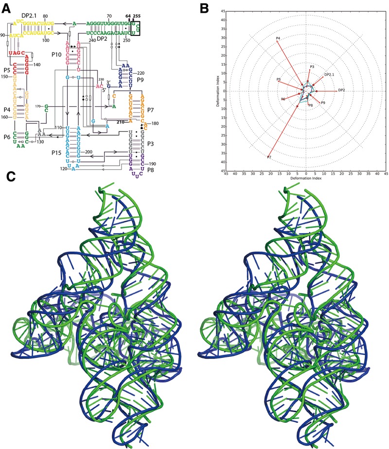
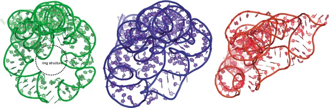
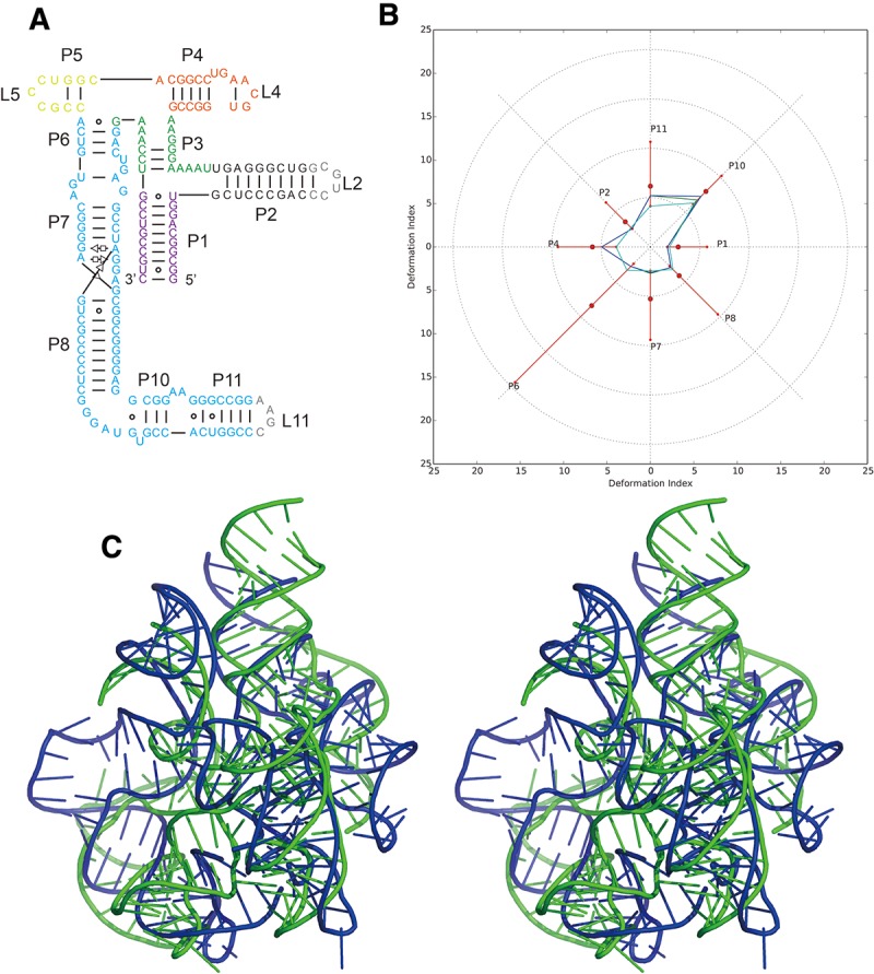
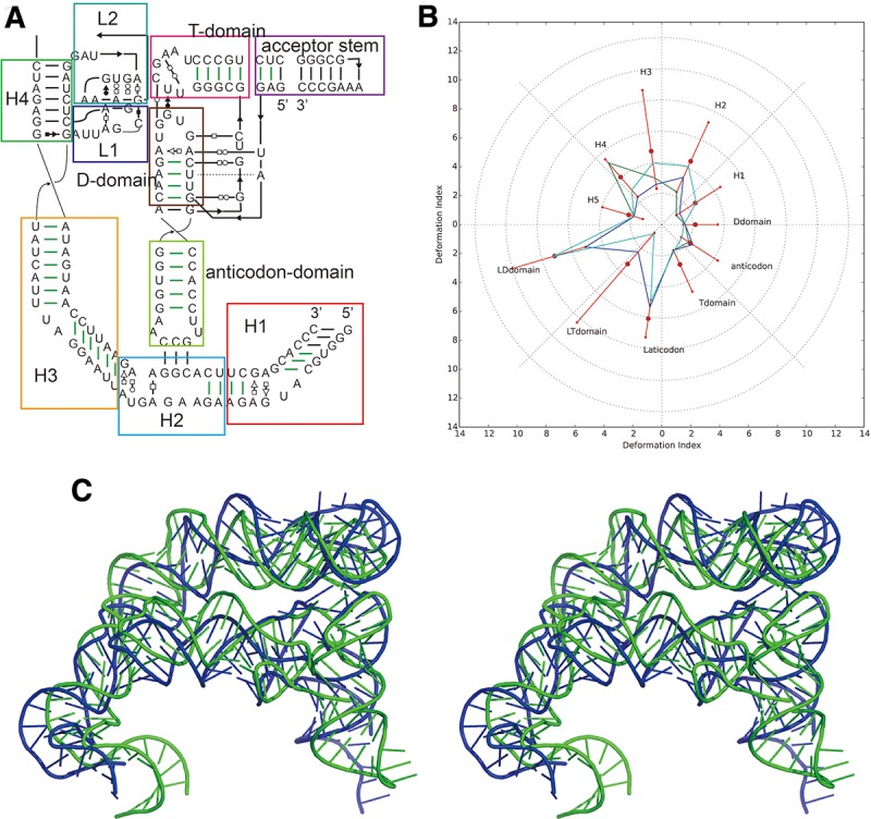
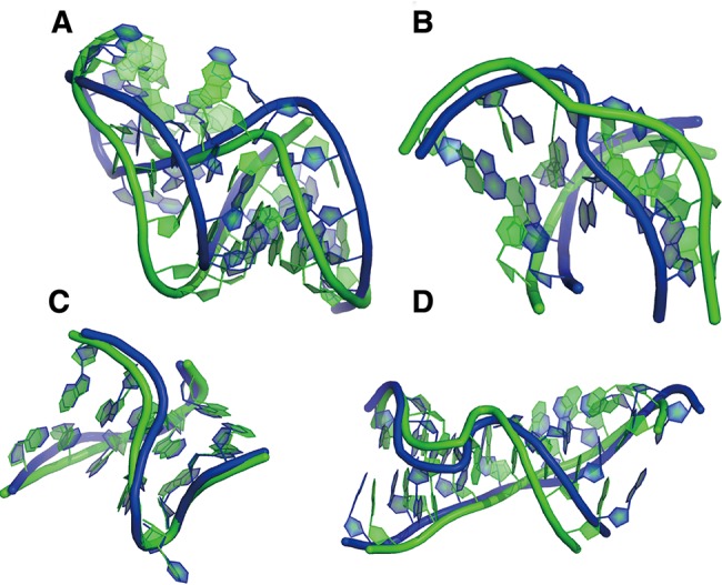
This paper is a report of a second round of RNA-Puzzles, a collective and blind experiment in three-dimensional (3D) RNA structure prediction. Three puzzles, Puzzles 5, 6, and 10, represented sequences of three large RNA structures with limited or no homology with previously solved RNA molecules. A lariat-capping ribozyme, as well as riboswitches complexed to adenosylcobalamin and tRNA, were predicted by seven groups using RNAComposer, ModeRNA/SimRNA, Vfold, Rosetta, DMD, MC-Fold, 3dRNA, and AMBER refinement. Some groups derived models using data from state-of-the-art chemical-mapping methods (SHAPE, DMS, CMCT, and mutate-and-map). The comparisons between the predictions and the three subsequently released crystallographic structures, solved at diffraction resolutions of 2.5-3.2 Å, were carried out automatically using various sets of quality indicators. The comparisons clearly demonstrate the state of present-day de novo prediction abilities as well as the limitations of these state-of-the-art methods. All of the best prediction models have similar topologies to the native structures, which suggests that computational methods for RNA structure prediction can already provide useful structural information for biological problems. However, the prediction accuracy for non-Watson-Crick interactions, key to proper folding of RNAs, is low and some predicted models had high Clash Scores. These two difficulties point to some of the continuing bottlenecks in RNA structure prediction. All submitted models are available for download at http://ahsoka.u-strasbg.fr/rnapuzzles/.
Keywords: 3D prediction; X-ray structures; bioinformatics; force fields; models; structure quality.
© 2015 Miao et al.; Published by Cold Spring Harbor Laboratory Press for the RNA Society.
Figures
Similar articles
-
RNA-Puzzles Round III: 3D RNA structure prediction of five riboswitches and one ribozyme.RNA. 2017 May;23(5):655-672. doi: 10.1261/rna.060368.116. Epub 2017 Jan 30. RNA. 2017. PMID: 28138060 Free PMC article.
-
RNA 3D Structure Modeling by Combination of Template-Based Method ModeRNA, Template-Free Folding with SimRNA, and Refinement with QRNAS.Methods Mol Biol. 2016;1490:217-35. doi: 10.1007/978-1-4939-6433-8_14. Methods Mol Biol. 2016. PMID: 27665602
-
Automated RNA 3D Structure Prediction with RNAComposer.Methods Mol Biol. 2016;1490:199-215. doi: 10.1007/978-1-4939-6433-8_13. Methods Mol Biol. 2016. PMID: 27665601
-
RNA Structure: Advances and Assessment of 3D Structure Prediction.Annu Rev Biophys. 2017 May 22;46:483-503. doi: 10.1146/annurev-biophys-070816-034125. Epub 2017 Mar 30. Annu Rev Biophys. 2017. PMID: 28375730 Review.
-
Computational modeling of RNA 3D structure based on experimental data.Biosci Rep. 2019 Feb 8;39(2):BSR20180430. doi: 10.1042/BSR20180430. Print 2019 Feb 28. Biosci Rep. 2019. PMID: 30670629 Free PMC article. Review.
Cited by
-
Structures to the people!Elife. 2015 Jul 8;4:e09249. doi: 10.7554/eLife.09249. Elife. 2015. PMID: 26153622 Free PMC article.
-
3D RNA and Functional Interactions from Evolutionary Couplings.Cell. 2016 May 5;165(4):963-75. doi: 10.1016/j.cell.2016.03.030. Epub 2016 Apr 14. Cell. 2016. PMID: 27087444 Free PMC article.
-
Predicting RNA distance-based contact maps by integrated deep learning on physics-inferred secondary structure and evolutionary-derived mutational coupling.Bioinformatics. 2022 Aug 10;38(16):3900-3910. doi: 10.1093/bioinformatics/btac421. Bioinformatics. 2022. PMID: 35751593 Free PMC article.
-
Limits in accuracy and a strategy of RNA structure prediction using experimental information.Nucleic Acids Res. 2019 Jun 20;47(11):5563-5572. doi: 10.1093/nar/gkz427. Nucleic Acids Res. 2019. PMID: 31106330 Free PMC article.
-
Molecular dynamics correctly models the unusual major conformation of the GAGU RNA internal loop and with NMR reveals an unusual minor conformation.RNA. 2018 May;24(5):656-672. doi: 10.1261/rna.064527.117. Epub 2018 Feb 6. RNA. 2018. PMID: 29434035 Free PMC article.
References
-
- Adams PL, Stahley MR, Kosek AB, Wang JM, Strobel SA 2004. Crystal structure of a self-splicing group I intron with both exons. Nature 430: 45–50. - PubMed
Publication types
MeSH terms
Substances
Grants and funding
LinkOut - more resources
Full Text Sources
Other Literature Sources