Review
. 2015 Jun;168(2):393-406.
doi: 10.1104/pp.15.00124. Epub 2015 Apr 16.

Vesicles versus Tubes: Is Endoplasmic Reticulum-Golgi Transport in Plants Fundamentally Different from Other Eukaryotes?

Affiliations
Review

Vesicles versus Tubes: Is Endoplasmic Reticulum-Golgi Transport in Plants Fundamentally Different from Other Eukaryotes?

David G Robinson et al. Plant Physiol. 2015 Jun.

Abstract

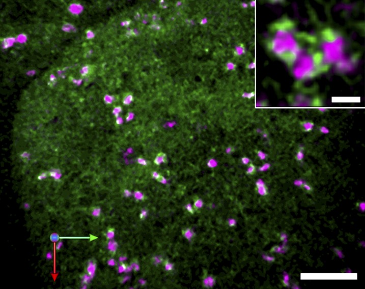
The endoplasmic reticulum (ER) is the gateway to the secretory pathway in all eukaryotic cells. Its products subsequently pass through the Golgi apparatus on the way to the cell surface (true secretion) or to the lytic compartment of the cell (vacuolar protein transport). In animal cells, the Golgi apparatus is present as a stationary larger order complex near the nucleus, and transport between the cortical ER and the Golgi complex occurs via an intermediate compartment which is transported on microtubules. By contrast, higher plant cells have discrete mobile Golgi stacks that move along the cortical ER, and the intermediate compartment is absent. Although many of the major molecular players involved in ER-Golgi trafficking in mammalian and yeast (Saccharomyces cerevisiae) cells have homologs in higher plants, the narrow interface (less than 500 nm) between the Golgi and the ER, together with the motility factor, makes the identification of the transport vectors responsible for bidirectional traffic between these two organelles much more difficult. Over the years, a controversy has arisen over the two major possibilities by which transfer can occur: through vesicles or direct tubular connections. In this article, four leading plant cell biologists attempted to resolve this issue. Unfortunately, their opinions are so divergent and often opposing that it was not possible to reach a consensus. Thus, we decided to let each tell his or her version individually. The review begins with an article by Federica Brandizzi that provides the necessary molecular background on coat protein complexes in relation to the so-called secretory units model for ER-Golgi transport in highly vacuolated plant cells. The second article, written by Chris Hawes, presents the evidence in favor of tubules. It is followed by an article from David Robinson defending the classical notion that transport occurs via vesicles. The last article, by Akihiko Nakano, introduces the reader to possible alternatives to vesicles or tubules, which are now emerging as a result of exciting new developments in high-resolution light microscopy in yeast.


Figures

Figure 1.
Figure 1.
Electron microscopy of COPII budding. A and B, Transitional ER plus adjacent Golgi stacks in the green alga C. noctigama as seen in chemically fixed (A) and high-pressure frozen samples (B). The cis-trans (c and t) polarity of the Golgi stacks is clearly visible and so too are budding and released COPII vesicles (arrowheads). Putative COPI vesicles are marked with arrows. C, High-pressure frozen endosperm cell of Arabidopsis. Budding COPII vesicles are marked with arrowheads, and free putative COPII vesicles are marked with arrows. D to G, Collage of COPII budding profiles. Note that many of the buds are at the termini of ER cisternae. Note that the ER in high-pressure frozen samples is, in general, much more dilated than in chemically fixed samples; in C. noctigama, it is extremely dilated (the ER in B can be recognized by the ribosomes at the left of the vacuole-like structure). Bars = 200 nm.
Figure 2.
Figure 2.
Golgi cisternae (rat sialyltransferase transmembrane domain and cytosolic tail fused to the yellow fluorescent protein, red) and the ERES marker (SEC16-GFP, green) visualized in tobacco leaf epidermal cells. Images from time-lapse sequence acquired at the cortical region of tobacco leaf epidermal cell with a Zeiss LSM510 confocal microscope. The Sec16 marker distributes at the peri-Golgi area (arrowheads) as well as to structures of unknown identity that are not associated with the Golgi marker (arrows; Takagi et al., 2013). The structures labeled by Sec16 can assume a ring-like shape (Takagi et al., 2013). Time of frames in the sequence is indicated at the left-hand corner of images (seconds). *, A chloroplast that is visible through chlorophyll autofluorescence. Bars = 5 and 1 μm (inset).
Figure 3.
Figure 3.
A, Maximum-intensity projection in negative contrast of a stack of thin sections from a tomogram of a pea (Pisum sativum) root tip Golgi body and associated ER impregnated by the osmium zinc iodide technique. The reconstruction is presented at an angle to show a clear tubular connection between the ER and cis-Golgi. B, Inside face view of a dry-cleaved carrot (Daucus carota) suspension culture cell. The cell had been fixed on a coated EM grid, dehydrated, and critical point dried prior to dry cleaving on double-sided tape. The view onto the plasma membrane shows dark mitochondria (M), complete Golgi stacks in face view (G), cisternal ER (CER), and tubular ER (arrows). Note the huge difference between the diameter of a Golgi body and ER tubules.
Figure 4.
Figure 4.
Organization of the ERESs in the budding yeast S. cerevisiae. Left, Dual-color three-dimensional image of Sec13-GFP (ERES marker, green) and monomeric red fluorescent protein-Sec12 (bulk ER marker, red) obtained by SCLIM. Right, Two-dimensional slice image taken from the three-dimensional data. ERESs localize at the high-curvature domains of the ER, such as along tubules and at the edge of the sheet. Bar = 1 μm.
Figure 5.
Figure 5.
The cis-Golgi cisternae (monomeric red fluorescent protein-SYP31, magenta) and ERESs (SEC13-yellow fluorescent protein, green) visualized in tobacco BY2 cells. Confocal images were captured by SCLIM and reconstructed into three dimensions with deconvolution. A typical trajectory image from a three-dimensional time-lapse movie is shown. Almost all of the Golgi stacks were associated with bright spots of ERESs. From the magnified image (inset), we could observe the ERESs surrounding the cis-Golgi making ring-shaped fluorescent patterns. Bars = 5 and 1 μm (inset).

References

    1. Andreeva AV, Kutuzov MA, Evans DE, Hawes CR (1998) The structure and function of the Golgi apparatus: a hundred years of questions. J Exp Bot 49: 1281–1291
    1. Appenzeller-Herzog C, Hauri HP (2006) The ER-Golgi intermediate compartment (ERGIC): in search of its identity and function. J Cell Sci 119: 2173–2183 - PubMed
    1. Bacia K, Futai E, Prinz S, Meister A, Daum S, Glatte D, Briggs JA, Schekman R (2011) Multibudded tubules formed by COPII on artificial liposomes. Sci Rep 1: 17. - PMC - PubMed
    1. Bar-Peled M, Raikhel NV (1997) Characterization of AtSEC12 and AtSAR1: Proteins likely involved in endoplasmic reticulum and Golgi transport. Plant Physiol 114: 315–324 - PMC - PubMed
    1. Barlowe C, Orci L, Yeung T, Hosobuchi M, Hamamoto S, Salama N, Rexach MF, Ravazzola M, Amherdt M, Schekman R (1994) COPII: a membrane coat formed by Sec proteins that drive vesicle budding from the endoplasmic reticulum. Cell 77: 895–907 - PubMed

Publication types

MeSH terms

LinkOut - more resources