

Protein structure. Direct observation of structure-function relationship in a nucleic acid-processing enzyme
- PMID: 25883359
- PMCID: PMC4424897
- DOI: 10.1126/science.aaa0130
Protein structure. Direct observation of structure-function relationship in a nucleic acid-processing enzyme
Abstract
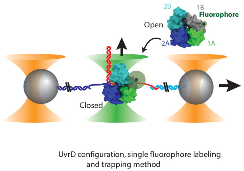
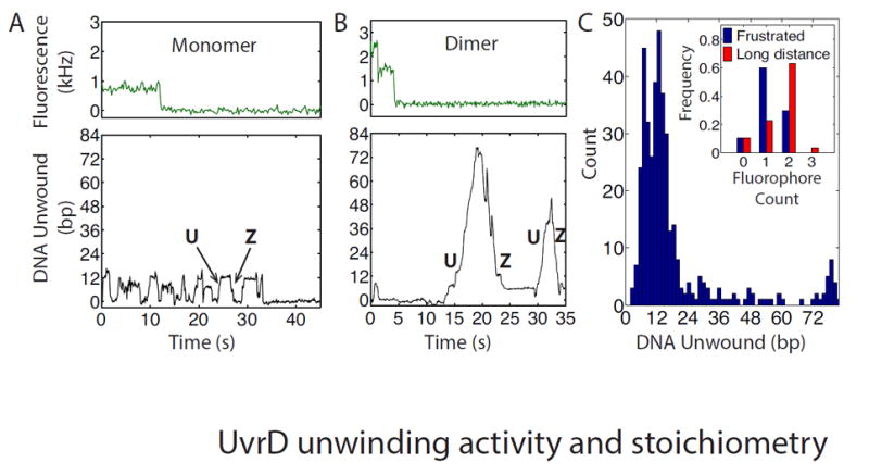
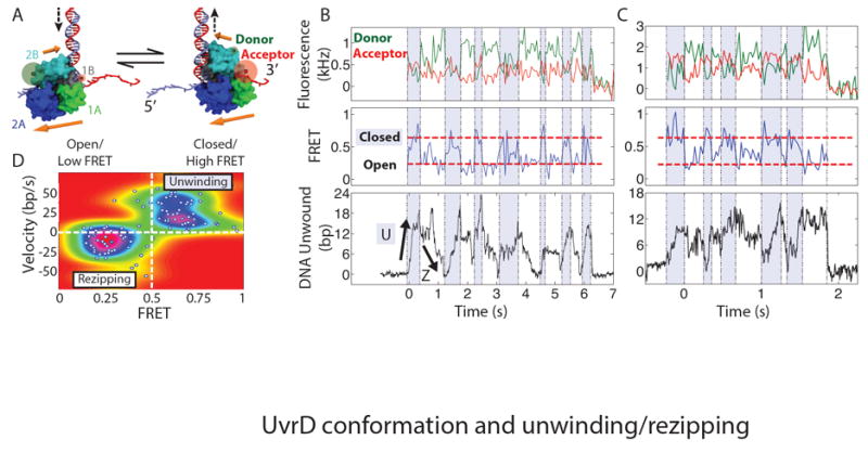
The relationship between protein three-dimensional structure and function is essential for mechanism determination. Unfortunately, most techniques do not provide a direct measurement of this relationship. Structural data are typically limited to static pictures, and function must be inferred. Conversely, functional assays usually provide little information on structural conformation. We developed a single-molecule technique combining optical tweezers and fluorescence microscopy that allows for both measurements simultaneously. Here we present measurements of UvrD, a DNA repair helicase, that directly and unambiguously reveal the connection between its structure and function. Our data reveal that UvrD exhibits two distinct types of unwinding activity regulated by its stoichiometry. Furthermore, two UvrD conformational states, termed "closed" and "open," correlate with movement toward or away from the DNA fork.
Copyright © 2015, American Association for the Advancement of Science.
Figures
Similar articles
-
Large domain movements upon UvrD dimerization and helicase activation.Proc Natl Acad Sci U S A. 2017 Nov 14;114(46):12178-12183. doi: 10.1073/pnas.1712882114. Epub 2017 Oct 30. Proc Natl Acad Sci U S A. 2017. PMID: 29087333 Free PMC article.
-
Direct Visualization of Helicase Dynamics Using Fluorescence Localization and Optical Trapping.Methods Enzymol. 2017;582:121-136. doi: 10.1016/bs.mie.2016.08.004. Epub 2016 Oct 27. Methods Enzymol. 2017. PMID: 28062032
-
UvrD helicase activation by MutL involves rotation of its 2B subdomain.Proc Natl Acad Sci U S A. 2019 Aug 13;116(33):16320-16325. doi: 10.1073/pnas.1905513116. Epub 2019 Jul 30. Proc Natl Acad Sci U S A. 2019. PMID: 31363055 Free PMC article.
-
Roles of the C-Terminal Amino Acids of Non-Hexameric Helicases: Insights from Escherichia coli UvrD.Int J Mol Sci. 2021 Jan 20;22(3):1018. doi: 10.3390/ijms22031018. Int J Mol Sci. 2021. PMID: 33498436 Free PMC article. Review.
-
Interplay between DNA replication, recombination and repair based on the structure of RecG helicase.Philos Trans R Soc Lond B Biol Sci. 2004 Jan 29;359(1441):49-59. doi: 10.1098/rstb.2003.1364. Philos Trans R Soc Lond B Biol Sci. 2004. PMID: 15065656 Free PMC article. Review.
Cited by
-
Chemo-mechanical pushing of proteins along single-stranded DNA.Proc Natl Acad Sci U S A. 2016 May 31;113(22):6194-9. doi: 10.1073/pnas.1602878113. Epub 2016 May 16. Proc Natl Acad Sci U S A. 2016. PMID: 27185951 Free PMC article.
-
Comprehensive Characterization of the Recombinant Catalytic Subunit of cAMP-Dependent Protein Kinase by Top-Down Mass Spectrometry.J Am Soc Mass Spectrom. 2019 Dec;30(12):2561-2570. doi: 10.1007/s13361-019-02341-0. Epub 2019 Dec 2. J Am Soc Mass Spectrom. 2019. PMID: 31792770 Free PMC article.
-
Alignment of helicases on single-stranded DNA increases activity.Methods Enzymol. 2022;672:29-54. doi: 10.1016/bs.mie.2022.03.066. Epub 2022 Apr 26. Methods Enzymol. 2022. PMID: 35934480 Free PMC article.
-
Alternative transcription cycle for bacterial RNA polymerase.Nat Commun. 2020 Jan 23;11(1):448. doi: 10.1038/s41467-019-14208-9. Nat Commun. 2020. PMID: 31974358 Free PMC article.
-
The mechanism of gap creation by a multifunctional nuclease during base excision repair.Sci Adv. 2021 Jul 14;7(29):eabg0076. doi: 10.1126/sciadv.abg0076. Print 2021 Jul. Sci Adv. 2021. PMID: 34261654 Free PMC article.
References
-
- Yamaguchi M, Dao V, Modrich P. MutS and MutL activate DNA helicase II in a mismatch-dependent manner. J Biol Chem. 1998;273:9197–9201. - PubMed
-
- Matson SW. Escherichia coli helicase II (urvD gene product) translocates unidirectionally in a 3’ to 5’ direction. J Biol Chem. 1986;261:10169–10175. - PubMed
-
- Fischer C, Maluf N, Lohman T. Mechanism of ATP-dependent translocation of E. coli UvrD monomers along single-stranded DNA. J Mol Biol. 2004;344:1287–1309. - PubMed
Publication types
MeSH terms
Substances
Grants and funding
LinkOut - more resources
Full Text Sources
Other Literature Sources
Molecular Biology Databases
