

Case Reports
doi: 10.4250/jcu.2015.23.1.48.
Epub 2015 Mar 30.
Double chambered right ventricle with ventricular septal defect in adults: case series and review of the literature
Affiliations
- PMID: 25883758
- PMCID: PMC4398786
- DOI: 10.4250/jcu.2015.23.1.48
Item in Clipboard
Case Reports
Double chambered right ventricle with ventricular septal defect in adults: case series and review of the literature
J Cardiovasc Ultrasound.
2015 Mar.
Abstract
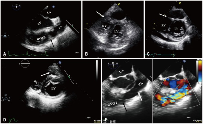
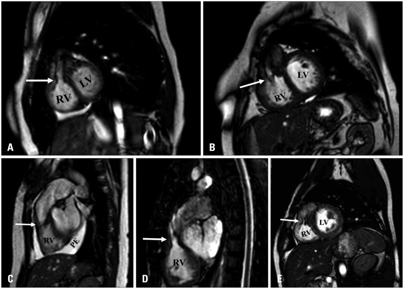
Double-chambered right ventricle (DCRV) is an uncommon congenital anomaly in which anomalous muscle bands divide the right ventricle into two chambers; a proximal high-pressure and distal low-pressure chamber. It may be associated with mid right ventricular obstruction. It is commonly associated with other congenital anomalies, most frequently perimembranous ventricular septal defect (PM-VSD). We herein present 5 adult patients with concomitant DCRV and PM-VSD who varied in their symptomatic presentations and the ways of management.
Keywords: Double chambered right ventricle; Echocardiography; Magnetic resonance; Ventricular septal defect.
Figures
References
-
- Wong PC, Sanders SP, Jonas RA, Colan SD, Parness IA, Geva T, Van Praagh R, Spevak PJ. Pulmonary valve-moderator band distance and association with development of double-chambered right ventricle. Am J Cardiol. 1991;68:1681–1686. - PubMed
-
- Hindle WV, Jr, Engle MA, Hagstrom JW. Anomalous right ventricular muscles: a clinicopathologic study. Am J Cardiol. 1968;21:487–495. - PubMed
-
- McElhinney DB, Chatterjee KM, Reddy VM. Double-chambered right ventricle presenting in adulthood. Ann Thorac Surg. 2000;70:124–127. - PubMed
-
- Nagashima M, Tomino T, Satoh H, Nakata T, Ohtani T, Saito H. Double-chambered right ventricle in adulthood. Asian Cardiovasc Thorac Ann. 2005;13:127–130. - PubMed
-
- Oliver JM, Garrido A, González A, Benito F, Mateos M, Aroca A, Sanz E. Rapid progression of midventricular obstruction in adults with double-chambered right ventricle. J Thorac Cardiovasc Surg. 2003;126:711–717. - PubMed
Publication types
LinkOut - more resources
Full Text Sources
Other Literature Sources
