. 2015:2015:458423.
doi: 10.1155/2015/458423. Epub 2015 Mar 25.

Metastatic colonic adenocarcinoma in breast: report of two cases and review of the literature

Affiliations

Metastatic colonic adenocarcinoma in breast: report of two cases and review of the literature

Jiten P Kothadia et al. Case Rep Oncol Med. 2015.

Abstract

Metastatic adenocarcinoma to the breast from an extramammary site is extremely rare. In the literature, the most current estimate is that extramammary metastases account for only 0.43% of all breast malignancies and that, of these extramammary sites, colon cancer metastases form a very small subset. Most commonly seen metastasis in breast is from a contralateral breast carcinoma, followed by metastasis from hematopoietic neoplasms, malignant melanoma, sarcoma, lung, prostate, and ovary and gastric neoplasms. Here we present two rare cases, in which colonic adenocarcinomas were found to metastasize to the breast. In both cases, core biopsies were obtained from the suspicious areas identified on mammogram. Histopathology revealed neoplastic proliferation of atypical glandular components within benign breast parenchyma which were morphologically consistent with metastatic adenocarcinoma. By immunohistochemical staining, it was confirmed that the neoplastic components were immunoreactive to colonic markers and nonreactive to breast markers, thus further supporting the morphologic findings. It is extremely important to make this distinction between primary breast cancer and a metastatic process, in order to provide the most effective and appropriate treatment for the patient and to avoid any harmful or unnecessary surgical procedures.


Figures
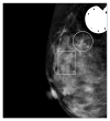
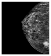
Figure 1
Figure 1
Mammographic image of the breast showing 2 groupings (A and B) of microcalcifications in the upper medial aspect of the right breast.
Figure 2
Figure 2
Mammographic image of the breast showing highly vascular spiculated hypoechoic mass measuring 1.35 cm × 1.46 cm × 1.22 cm (arrow).
Figure 3
Figure 3
Ultrasonography image of the right breast showing 1.4 cm × 1.2 cm irregularly shaped, speculated, hypoechoic lesion with central necrotic changes and high vascularity.
Figure 4
Figure 4
Tissue core biopsy from suspicious areas shows benign appearing breast terminal ductal-lobular units (TDLU) (blue arrows), interspersed by neoplastic colonic glands (black arrows) without any in situ carcinoma component (Hematoxylin and Eosin stain, 40x magnification).
Figure 5
Figure 5
Neoplastic colonic glands (A and B) show hyperchromatic, crowded nuclei, loss of nuclear polarity, and pseudostratification, intermixed with normal breast ducts (C). Necrosis is noted in neoplastic colonic glands (arrow) (Hematoxylin and Eosin stain, 200x magnification).
Figure 6
Figure 6
Metastatic colonic glands show intraglandular microcalcifications (arrow) (Hematoxylin and Eosin stain, 200x magnification).
Figure 7
Figure 7
Cytokeratin 20 immunostaining in both cases shows positive cytoplasmic staining in metastatic colonic glands (200x magnification).
Figure 8
Figure 8
CDX-2 immunostaining in both cases shows intense positivity in nucleus of neoplastic colonic glands (200x magnification).
Figure 9
Figure 9
In both of these cases, benign breast ductal cells are immunoreactive to estrogen receptor (left) and nonreactive to metastatic colonic glands (right) (200x magnification).

References

    1. Selcukbiricik F., Tural D., Bay A., Sahingoz G., Ilvan S., Mandel N. M. A malignant mass in the breast is not always breast cancer. Case Reports in Oncology. 2011;4(3):521–525. doi: 10.1159/000334079. - DOI - PMC - PubMed
    1. Cabibi D., Cipolla C., Valerio M. R., Ingrao S. Metastatic seeding of colon adenocarcinoma manifesting as synchronous breast and chest wall localization: report of a case. Surgery Today. 2011;41(2):242–246. doi: 10.1007/s00595-009-4222-3. - DOI - PubMed
    1. Perin T., Canzonieri V., Memeo L., Massarut S. Breast metastasis of primary colon cancer with micrometastasis in the axillary sentinel node: a metastasis that metastasized? Diagnostic Pathology. 2011;6, article 45 doi: 10.1186/1746-1596-6-45. - DOI - PMC - PubMed
    1. de Bobadilla L. F., Villanueva A. G., Collado M., et al. Breast metastasis of primary colon cancer. Revista Española de Enfermedades Digestivas. 2004;96(6):415–419. - PubMed
    1. Georgiannos S. N., Chin Aleong J., Goode A. W., Sheaff M. Secondary neoplasms of the breast: a survey of the 20th Century. Cancer. 2001;92(9):2259–2266. doi: 10.1002/1097-0142(20011101)92:9x003C;2259::aid-cncr1571x0003e;3.0.co;2-o. - DOI - PubMed

LinkOut - more resources