Review
. 2015 Apr 1:3:21.
doi: 10.3389/fcell.2015.00021. eCollection 2015.

Atypical centrioles during sexual reproduction

Affiliations
Review

Atypical centrioles during sexual reproduction

Tomer Avidor-Reiss et al. Front Cell Dev Biol. .

Abstract

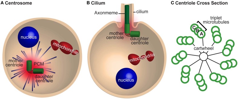
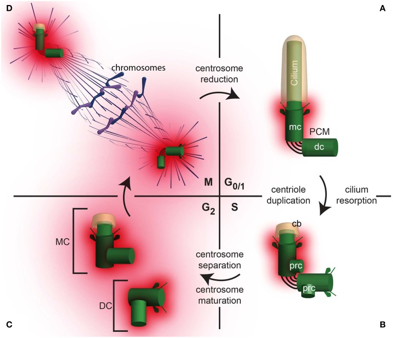
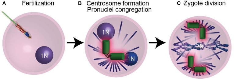
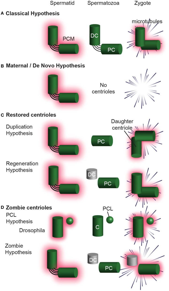
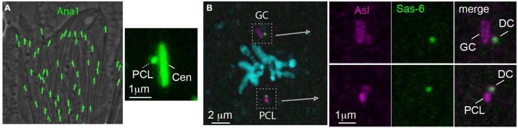
Centrioles are conserved, self-replicating, microtubule-based, 9-fold symmetric subcellular organelles that are essential for proper cell division and function. Most cells have two centrioles and maintaining this number of centrioles is important for animal development and physiology. However, how animals gain their first two centrioles during reproduction is only partially understood. It is well established that in most animals, the centrioles are contributed to the zygote by the sperm. However, in humans and many animals, the sperm centrioles are modified in their structure and protein composition, or they appear to be missing altogether. In these animals, the origin of the first centrioles is not clear. Here, we review various hypotheses on how centrioles are gained during reproduction and describe specialized functions of the zygotic centrioles. In particular, we discuss a new and atypical centriole found in sperm and zygote, called the proximal centriole-like structure (PCL). We also discuss another type of atypical centriole, the "zombie" centriole, which is degenerated but functional. Together, the presence of centrioles, PCL, and zombie centrioles suggests a universal mechanism of centriole inheritance among animals and new causes of infertility. Since the atypical centrioles of sperm and zygote share similar functions with typical centrioles in somatic cells, they can provide unmatched insight into centriole biology.

Keywords: centriole; centrosome; cilium; fertilization; microtubules; reproduction; sperm; zygote.


Figures

Figure 1
Figure 1
The centrosome is required for mitosis and cilia nucleation. (A) A cell with a centrosome emanating astral microtubules (blue lines). The mother and daughter centrioles are tethered together and are surrounded by pericentriolar material (PCM). (B) A cell nucleating a cilium. The mother centriole nucleates the axoneme and has less PCM and astral microtubules than in (A). The centrioles are still tethered together. (C) A cross section of a centriole with a cartwheel and triplet microtubules with 9-fold symmetric. The microtubules in the triplet are referred to as tubules (A–C) depending on their position.
Figure 2
Figure 2
The centrosome changes throughout the cell cycle. (A) A daughter cell inherits one centrosome, which then undergoes centrosome reduction, where the PCM quantity is reduced and the centrosome migrates to the cell periphery where it nucleate a cilium in G0/1. In G0/1, the centrioles (mc, mother centriole; dc, daughter centriole) are tethered together, with PCM surrounding the mother centriole. (B) In S phase, the cilium is reabsorbed, leaving a ciliary bud (cb). Coincident with DNA replication in the nucleus, procentrioles (prc) form, each one orthogonal to the mother or daughter centriole, respectively. (C) In G2, the tether between the mother and daughter centriole is severed, allowing the centrosomes to separate. The centrosome containing the original mother centriole is often called the Mother Centrosome (MC), whereas the original daughter centriole is now part of the Daughter Centrosome (DC). Both the Mother Centrosome and Daughter Centrosome recruit more PCM. (D) In M phase, the centrosomes migrate to opposite poles of the cell and regulate chromosome segregation.
Figure 3
Figure 3
Four groups of centrosomal proteins. (A) The cascade of proteins required for initiating the formation of the procentriole (prc) near the proximal end of the mother centriole (mc). (B) A quarter of a cross-section of a centriole, showing cartwheel and microtubule wall proteins. (C) A quarter of a cross-section of a centriole, showing a matrix of PCM proteins. Adapted from Mennella et al. (2012). (D) Transition zone (tz), subdistal appendages, and distal centriole proteins that are required for cilium formation.
Figure 4
Figure 4
The centrioles are essential for pronucleus migration. (A) After the sperm fertilizes the ovum, it provides the zygote with modified centrioles and its genetic material in the form of a male pronucleus. (B) The centrioles (green) then recruit maternal PCM proteins (pink) and nucleate astral microtubules (purple lines), which mediate the migration of pronuclei (blue spheres) using motor proteins. (C) Following duplication, the centrosomes found in the spindle poles mediate chromosome segregation. N and 2N indicate chromosome copy number.
Figure 5
Figure 5
The centrosome is eliminated during oogenesis. (A) During meiosis I in humans, the 4N primary oocyte divides and the centrioles degrade during prophase I. (B) The resulting secondary oocyte then undergoes meiosis II in the absents centrioles. (C) The culmination of oogenesis is an ovum with an unduplicated genome (1N), which lacks centrioles. Mt, mitochondria. N and 2N indicate chromosome copy number.
Figure 6
Figure 6
The centrosome is reduced during spermatogenesis. (A) Mammalian spermatogenesis: In the primary spermatocyte, the two centrioles duplicate as the DNA replicates. After meiosis I, the secondary spermatocyte with two centrioles duplicates, while the DNA does not. For this reason, Meiosis II culminates with two centrioles in each of the spermatids. The spermatid then undergoes spermiogenesis, during which the distal centriole forms a flagellum, and the centrosome is reduced. Human ejaculated spermatozoa have one intact centriole and one degenerated centriole. (B) Drosophila spermatogenesis: Spermatogenesis in flies. Note that during meiosis I and meiosis II the centrioles do not duplicate, and each spermatid has only one centriole (c). This centriole then buds off the PCL and gives rise to a flagellum. Both the centriole and PCL loses most of their component proteins, as part of centrosome reduction. PC, proximal centriole; DC, distal centriole; Mt, mitochondria. N and 2N indicate chromosome copy number.
Figure 7
Figure 7
Models for centriole number in sperm vary from organism to organism. A phylogenetic tree with the varied centriole numbers in different organisms. Organisms with two intact centrioles in the spermatozoa (black). Organisms with one centriole and one PCL (red). Organisms with no centrioles in spermatozoa (green). Organisms with one centriole and one degenerated centriole in spermatozoa (blue).
Figure 8
Figure 8
Centrosome reduction occurs in three steps. (A) Centrosome reduction in humans. The centrosome that has the future distal centriole (DC) and proximal centriole (PC) first loses its ability to nucleate astral microtubules. Next, it loses its PCM, and then finally, its centriolar proteins, leaving one centriole with intact microtubular structures and one degenerated. (B) In Drosophila, centrosome reduction occurs in the same three steps. Both the PCL and the centriole then undergo reduction, losing most, but not all of their comprising proteins, and the centriole degenerates. (C) An EM picture of the human proximal centriole (PC) in the connecting piece of spermatozoa, showing clear triplet microtubules with 9 fold symmetry. Obtained with permission from Rawe et al. (2008). (D,E) In Drosophila melanogaster, the PCL and centriole (C) undergo centrosome reduction (Blachon et al., 2014). (D) The centriole (C, white solid line) is intensely labeled by PACT-GFP (which is over-expressed by the strong ubiquitin promoter in intermediate spermatids), but is barely observed at the base of the sperm nucleus (see inset for magnification of this giant centriole). (E) The PCL (white dashed line) and centriole (C, white solid line) are observed in intermediate spermatids by Ana1-GFP. However, Ana1-GFP is not observed in spermatozoa found in the seminal vesicle (white arrowhead). Scalebar 1 μm (Blachon et al., 2014).
Figure 9
Figure 9
Hypotheses for centriole Inheritance. (A) The classical hypothesis, where, after centrosome reduction, two centrioles are inherited, and they form the zygote centrosome. (B) The maternal/de novo hypothesis, in which the spermatid has two centrioles, which are then eliminated in the spermatozoa. The zygote then undergoes division without centrioles. (C) Hypotheses that involve centrioles degenerating and then being restored to their original configuration upon fertilization. In the Duplication Hypothesis, one centriole is eliminated, and the second centriole is restored using the remaining centriole as a template. The Regeneration Hypothesis suggests that the distal centriole partially degenerates, but is restored upon fertilization. In this hypothesis, the centriole is only functional after restoration. (D) The Zombie Hypotheses state that degenerated centrioles may still be functional. For example, in mammals, the degenerated distal centriole is inherited into the zygote and functions without being restored. Similarly, the PCL is partially degenerated during spermiogenesis, but is inherited into the zygote and is functional. DC, distal centriole; PC, proximal centriole, C, centriole.
Figure 10
Figure 10
The PCL is a centriole precursor that forms in the spermatid and becomes one of the zygote's spindle poles. (A) Ana1-GFP labels the PCL that forms near the Ana-1 labeled giant centriole (GC) in Drosophila spermatids (Blachon et al., 2009). (B) A metaphase zygote formed from an ovum expressing Sas-6-GFP has both a PCL and GC centrosome (which are labeled by antibody against the PCM marker Asl). The PCL and GC centrosomes each have their own daughter centrioles (DC) (which are labeled by the maternal Sas-6- GFP) (Blachon et al., 2014).

Similar articles

Cited by

References

    1. Avasthi P., Marshall W. F. (2012). Stages of ciliogenesis and regulation of ciliary length. Differentiation 83, S30–S42. 10.1016/j.diff.2011.11.015 - DOI - PMC - PubMed
    1. Avidor-Reiss T., Gopalakrishnan J. (2013). Building a centriole. Curr. Opin. Cell Biol. 25, 72–77. 10.1016/j.ceb.2012.10.016 - DOI - PMC - PubMed
    1. Avidor-Reiss T., Gopalakrishnan J., Blachon S., Polyanovsky A. (2012). Centriole duplication and inheritance in Drosophila melanogaster, in The Centrosome: Cell and Molecular Mechanisms of Functions and Dysfunctions in Disease, ed Schatten H. (New York, NY: Humana Press; ), 3–31.
    1. Baccetti B., Afzelius B. A. (1976). The biology of the sperm cell. Monogr. Dev. Biol. 1–254. - PubMed
    1. Baccetti B., Burrini A. G., Dallai R., Pallini V. P., Periti P., Piantelli F., et al. . (1973). Structure and function in the spermatozoon of Bacillus rossius. The spermatozoon of arthropoda. XIX. J. Ultrastruct. Res. 12, 1–73. - PubMed

LinkOut - more resources