

Polymorphonuclear leukocyte apoptosis is accelerated by sulfatides or sulfatides-treated Salmonella Typhimurium bacteria
- PMID: 25883957
- PMCID: PMC4391312
- DOI: 10.1155/2015/381232
Polymorphonuclear leukocyte apoptosis is accelerated by sulfatides or sulfatides-treated Salmonella Typhimurium bacteria
Abstract
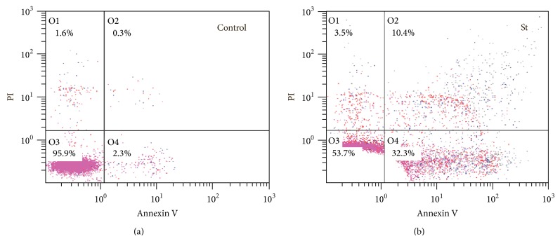
Neutrophils die by apoptosis following activation and uptake of microbes or enter apoptosis spontaneously at the end of their lifespan if they do not encounter a pathogen. Here we report that sulfatides or sulfatides-treated Salmonella Typhimurium bacteria accelerated human neutrophil apoptosis. Neutrophil apoptosis was examined by flow cytometry. Sulfatides caused prominent increase in percentage of apoptotic cells after 2.5 hrs of incubation. Salmonella Typhimurium bacteria by themselves did not affect the basal level of apoptosis in neutrophil population. When neutrophils were added to S. Typhimurium "opsonized" by sulfatides, apoptotic index significantly increased, whereas the number of phagocyting cells was not influenced. Sulfatides' proapoptotic effect was strongly dependent on the activity of β-galactosidase; inhibition of this enzyme impaired its potency to accelerate apoptosis. These data support the mechanism of neutrophil apoptosis triggering based on sulfatides' ability to accumulate in intracellular compartments and mediate successive increase in ceramide content resulting from β-galactosidase activity.
Figures


Similar articles
-
Sulfatides inhibit leukotriene synthesis in human polymorphonuclear granulocytes by a mechanism involving lipid rearrangement in intracellular membranes.Int J Biochem Cell Biol. 2008;40(1):110-24. doi: 10.1016/j.biocel.2007.07.001. Epub 2007 Jul 18. Int J Biochem Cell Biol. 2008. PMID: 17822942
-
De novo C16- and C24-ceramide generation contributes to spontaneous neutrophil apoptosis.J Leukoc Biol. 2007 Jun;81(6):1477-86. doi: 10.1189/jlb.0806529. Epub 2007 Feb 28. J Leukoc Biol. 2007. PMID: 17329567
-
Nitric oxide mediates distinct effects of various LPS chemotypes on phagocytosis and leukotriene synthesis in human neutrophils.Int J Biochem Cell Biol. 2010 Jun;42(6):921-31. doi: 10.1016/j.biocel.2010.01.025. Epub 2010 Feb 1. Int J Biochem Cell Biol. 2010. PMID: 20117233
-
Clustering of death receptors in lipid rafts initiates neutrophil spontaneous apoptosis.Biochem Soc Trans. 2004 Nov;32(Pt 5):679-81. doi: 10.1042/BST0320679. Biochem Soc Trans. 2004. PMID: 15493986 Review.
-
Neutrophil granulocytes as host cells and transport vehicles for intracellular pathogens: apoptosis as infection-promoting factor.Immunobiology. 2008;213(3-4):183-91. doi: 10.1016/j.imbio.2007.11.010. Epub 2008 Feb 8. Immunobiology. 2008. PMID: 18406366 Review.
Cited by
-
Type II NKT cells: a distinct CD1d-restricted immune regulatory NKT cell subset.Immunogenetics. 2016 Aug;68(8):665-76. doi: 10.1007/s00251-016-0930-1. Epub 2016 Jul 12. Immunogenetics. 2016. PMID: 27405300 Free PMC article. Review.
-
Adjunctive inhaled amikacin in infants with Ventilator-Associated Pneumonia optimizes the complex antimicrobial therapy: pilot study.Acta Biomed. 2023 Apr 24;94(2):e2023084. doi: 10.23750/abm.v94i2.13910. Acta Biomed. 2023. PMID: 37092633 Free PMC article.
-
Tumor ratio of unsaturated to saturated sulfatide species is associated with disease-free survival in intrahepatic cholangiocarcinoma.Cell Oncol (Dordr). 2023 Jun;46(3):629-642. doi: 10.1007/s13402-022-00766-6. Epub 2023 Jan 11. Cell Oncol (Dordr). 2023. PMID: 36630049 Free PMC article.
References
-
- Watson R. W. G., Redmond H. P., Wang J. H., Condron C., Bouchier-Hayes D. Neutrophils undergo apoptosis following ingestion of Escherichia coli . The Journal of Immunology. 1996;156(10):3986–3992. - PubMed
Publication types
MeSH terms
Substances
LinkOut - more resources
Full Text Sources
Other Literature Sources
