. 2015 Apr 17;13(4):e1002125.
doi: 10.1371/journal.pbio.1002125. eCollection 2015 Apr.

Dynamic endothelial cell rearrangements drive developmental vessel regression

Affiliations

Dynamic endothelial cell rearrangements drive developmental vessel regression

Claudio A Franco et al. PLoS Biol. .

Erratum in

Abstract

Patterning of functional blood vessel networks is achieved by pruning of superfluous connections. The cellular and molecular principles of vessel regression are poorly understood. Here we show that regression is mediated by dynamic and polarized migration of endothelial cells, representing anastomosis in reverse. Establishing and analyzing the first axial polarity map of all endothelial cells in a remodeling vascular network, we propose that balanced movement of cells maintains the primitive plexus under low shear conditions in a metastable dynamic state. We predict that flow-induced polarized migration of endothelial cells breaks symmetry and leads to stabilization of high flow/shear segments and regression of adjacent low flow/shear segments.


Conflict of interest statement

The authors have declared that no competing interests exist.

Figures
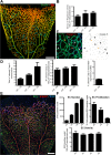
Fig 1
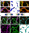
Fig 1. Developmental vessel regression does not depend on endothelial cell death.
A, Overview of a wild-type postnatal day 6 (P6) mouse retina highlighting all regression profiles (blue lines). Regression profiles are vessel segments with collagen IV-positive vessel segments and negative for IsolectinB4. B, Quantification of number of regressing segments at P4, P6, and P8 retinas per vascularized area. C, Representative image of a P6 mouse retina labeled with Col.IV (green), cleaved caspase-3 (red) and IsolectinB4 (blue) showing regression profiles (white arrows) associated with cleaved caspase-3-positive cells (yellow arrows). D, Quantification of total numbers of cleaved caspase-3 events in entire P4, P6, and P8 mouse retinas, normalized for 100 μm2 of vascularized tissue. At P6, only 4.82% ± 0.76 (n = 5 retinas) of regression events are associated with caspase-3-positive labeled endothelial cells. Data given as mean ± SD. E, Confocal images of P6 wild-type retinas after 4h EdU-treatment (EdU, blue), endothelial cell nuclei (Erg, green) and blood vessels (ICAM2, red). F, Quantification of total number of endothelial cells, percentage of ETS related gene (Erg)- and 5-ethynyl-2'-deoxyuridine (EdU)-positive cells to total number of endothelial cells, and number of endothelial cells per vascularized area at specified mouse retina developmental stages. Mean ± SEM; n = 4 mice, 2 litters. Scale bars (A and E: 200 μm; C: 25 μm). The data used to make this figure can be found in S1 Data.
Fig 2
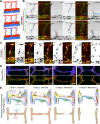
Fig 2. Developmental vessel regression resembles anastomosis in reverse.
A–C, Immunostaining for lumen (ICAM2), junctions (ZO1 and Cdh5), blood vessels (IB4), and basement membrane (Col.IV) shows that lumen breakage (arrows in A) and junction disconnection (arrows in B and C) is an early step in vessel regression. D and E, Single-cell labeling using Cre-induced expression of membrane-bound GFP (mGFP) shows polarized morphology of activated endothelial cells with filopodia projections (yellow arrows in in D and E). Endothelial cells (green dotted-lines) bridge two or more vessel segments in the regressing vessel, showing rings or points of junctional connection (blue arrows in in D and E). Scale bars (A–E: 10 μm).
Fig 3
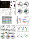
Fig 3. Disorganized endothelial cell polarity correlates with vessel regression.
A and B, Polarization of endothelial cells in a wild type (WT) P6 retina vascular network labeled for endothelial cell nuclei (Erg), basement membrane (Col.IV) lumen (intercellular adhesion molecule 2 [ICAM2]), and Golgi (130 kDa cis-Golgi matrix protein [GM130] or Golgi integral membrane protein 4 [Golph4]). Distance from the center of mass of each endothelial nucleus to the corresponding Golgi is used to draw a yellow (A) or pink (B) arrow, indicative of front–rear (axial) polarity. A, In anastomosis, endothelial polarities point towards each other. In regression, endothelial cells polarities point towards the neighboring vessel segments. Images were segmented for visualization purposes; original images can be found in S4 Fig B, Representative image of stochastic cell labeling using inducible Cre-lox mediated expression of membrane-bound GFP (mGFP), revealing the morphology of single endothelial cells in regressing vessels (white arrowhead) in combination with the axial polarity assessment (pink arrows). The white dotted line outlines one cell in a regression profile (lacking ICAM2), which is in contact with multiple vessel segments and shows an activated morphology with numerous filopodia. Scale bars (A and B: 10 μm).
Fig 4
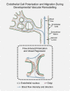
Fig 4. Extensive cell rearrangements drive developmental vessel regression.
A, Schematic of ISV disconnection from the aorta. B, S4 Movie still images from time-lapse confocal imaging at 48 h post-fertilization of a transgenic Tg(kdrl:mCherry-CAAX) zebrafish embryo injected with pTol2:fli1ep:eGFP-CAAX, showing the dynamic behavior of endothelial cells during the process of intersegmental vessel regression (white arrow), triggered by the anastomosis of a venous sprout (blue arrow). C, S5 Movie still images from time-lapse confocal imaging at 48 h post-fertilization of a transgenic Tg(Fli1a:dsRedEx); Tg(Fli1a:nEGFP) zebrafish embryo showing the dynamic behavior of endothelial cell nuclei during vessel regression (white arrow). The regressing endothelial cell (asterisk) is viable and undergoes mitosis a later stage, originating two daughter endothelial cells (asterisk a and b). D, Confocal images of regression profiles in a wild-type P6 mouse retina labeled with lumen (ICAM2), junctions (ZO1), and basement membrane (Col.IV). Vessel segments range from a normal stable vessel segment (left panel), stenosis lumen/junction, disconnected lumen, and complete absence of lumen (right panel). E, Proposed four-step model for vessel regression. Step 1: selection of the regressing branch, Step 2: lumen stenosis in the regressing vessel, Step 3: junction/lumen remodeling during endothelial cell retraction, and Step 4: integration of regressing endothelial cells in neighboring vessel segments leaving an empty basement membrane. (dlav: dorsolateral anastomotic vessel; isv: intersegmental vessel; da: dorsal aorta; pcv: posterior cardinal vein). Scale bars (B and C: 20 μm; D: 10 μm).
Fig 5
Fig 5. Coordinated polarity induced by high flow triggers vessel regression.
A, Overview of the axial polarization pattern of endothelial cells in a WT P6 retina vascular network labeled for endothelial cell nuclei (Erg), lumen (ICAM2), and Golgi (Golph4), and corresponding image segmentation of the vascular plexus in (a), highlighting the lumen of blood vessels (grey), and the axial polarity of all endothelial cells (red arrows). B, Analysis of the endothelial axial polarity angle in the main vessels, correlated to predicted blood flow direction by the rheology in silico model. Endothelial cells robustly position their Golgi apparatus against the blood flow in all vascular regions analyzed. C, Quantitative analysis of the percentage of endothelial cells polarized at 180°(±45°) compared to the flow direction in the different vascular beds (n = 3 retinas). D, Quantitative analysis of cell density, mean wall shear stress and branching point density in P6 mouse retina vascular plexus (n = 3 retinas). E, S6 Movie still images from time-lapse live imaging of a Tg(fli1a:eGFP) zebrafish embryo (grey) injected with pTol2:fli1ep:mCherry-GM130 (green). F, Quantification of endothelial cell axial polarity in ISVs showing dorsal axial polarization during the sprouting phase (not lumenized). In stable ISVs, endothelial shows significantly enriched dorsal or ventral axial polarity when in venous or arterial ISVs, respectively, corresponding to polarization against the predicted blood flow direction. G, Representative images of axial polarity and color-coded representation of the rheology prediction for velocity and wall shear stress in the corresponding vessel segments. Axial polarity length correlates with higher levels of luminal shear stress. In low shear vessels endothelial cells show decreased polarization and tend to point towards high flow vessel segments (black arrows). Scale bars (A: 50 μm; E: 20 μm). The data used to make this figure can be found in S1 Data.
Fig 6
Fig 6. Working model for flow-induced remodeling through directional migration.
Schematized prototypic vessel network in a developing retina. Endothelial cell axial polarity is indicated by Golgi position; flow direction (arrows) and velocity (thickness), producing luminal membrane shear stress, are depicted by light blue lines. Vessels in the distal primitive plexus are exposed to low, oscillatory, or no-flow, and vessels closer to developing arteries are exposed to higher blood flow velocities. High blood flow leads to increased levels of shear stress, which induces robust polarization of endothelial cells against flow. Increasing flow asymmetries between juxtaposed vessel segments trigger endothelial migration away from low flow regions (black arrows), inducing vessel segment regression.

References

    1. Potente M, Gerhardt H, Carmeliet P (2011) Basic and therapeutic aspects of angiogenesis. Cell 146: 873–887. 10.1016/j.cell.2011.08.039 - DOI - PubMed
    1. Carmeliet P (2005) Angiogenesis in life, disease and medicine. Nature 438: 932–936. - PubMed
    1. Lobov IB, Rao S, Carroll TJ, Vallance JE, Ito M, et al. (2005) WNT7b mediates macrophage-induced programmed cell death in patterning of the vasculature. Nature 437: 417–421. - PMC - PubMed
    1. Meeson AP, Argilla M, Ko K, Witte L, Lang RA (1999) VEGF deprivation-induced apoptosis is a component of programmed capillary regression. Development 126: 1407–1415. - PubMed
    1. Hughes S, Chang-Ling T (2000) Roles of endothelial cell migration and apoptosis in vascular remodeling during development of the central nervous system. Microcirculation 7: 317–333. - PubMed

Publication types