

Intravitreal cilium associated with retinal detachment 40 years following penetrating eye injury: a case report
- PMID: 25884640
- PMCID: PMC4367883
- DOI: 10.1186/s12886-015-0010-6
Intravitreal cilium associated with retinal detachment 40 years following penetrating eye injury: a case report
Abstract
Background: The presence of an intraocular cilium is very rare and the response of the eye to the cilium is variable. We present the case of a patient with a cilium found in the vitreous cavity during vitrectomy for rhegmatogenous retinal detachment 40 years following penetrating eye injury. To our knowledge, this is the longest reported presence of a cilium in the vitreous cavity.
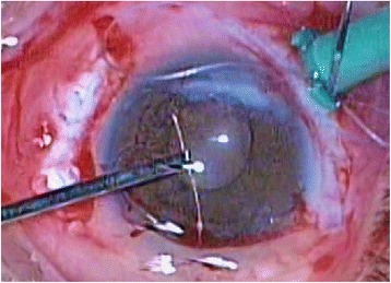
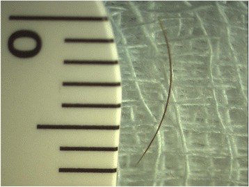
Case presentation: A 70-year-old Caucasian woman presented to the emergency department of our hospital complaining of sudden visual impairment and floaters of her right eye initiated 2 weeks earlier. Ophthalmic history included a penetrating injury of the right eye with a sharp metallic object 40 years ago and an uncomplicated phacoemulsification surgery in the same eye 2 years earlier. Fundoscopy revealed an inferior macula off rhegmatogenous retinal detachment. No inflammation was present. During vitrectomy and under scleral indentation at 5-o'clock position, a cilium was found at far retinal periphery. One end of the cilium was embedded in the retina, whereas the other end floated freely in the vitreous. The cilium was removed through the pars plana sclerotomy with intraocular foreign body forceps. The procedure was completed without any complications.
Conclusion: Penetrating eye injury is the most possible cause of cilium entrance in vitreous cavity in this case, which suggests that cilium can be well tolerated in vitreous cavity for as long as 40 years.
Figures

References
-
- Orhan M, Tatlipinar S, Irkec M. Corneal graft rejection and recurrent anterior uveitis associated with intraocular cilium. Ophthalmic Surg Lasers. 2002;33:231–2. - PubMed
-
- Oh KT, Oh KT, Singerman LJ. An eyelash in the vitreous cavity without apparent etiology. Ophthalmic Surg Lasers. 1996;27:243–5. - PubMed
Publication types
MeSH terms
LinkOut - more resources
Full Text Sources
Other Literature Sources
Miscellaneous
