. 2015 Mar 26:8:21.
doi: 10.1186/s13041-015-0111-4.

Ca(2+)/calmodulin-dependent protein kinase II interacts with group I metabotropic glutamate and facilitates receptor endocytosis and ERK1/2 signaling: role of β-amyloid

Affiliations

Ca(2+)/calmodulin-dependent protein kinase II interacts with group I metabotropic glutamate and facilitates receptor endocytosis and ERK1/2 signaling: role of β-amyloid

Fitore Raka et al. Mol Brain. .

Abstract

Background: Agonist stimulation of Group I metabotropic glutamate receptors (mGluRs) initiates their coupling to the heterotrimeric G protein, Gαq/11, resulting in the activation of phospholipase C, the release of Ca(2+) from intracellular stores and the subsequent activation of protein kinase C. However, it is now recognized that mGluR5a also functions as a receptor for cellular prion protein (PrP(C)) and β-amyloid peptide (Aβ42) oligomers to facilitate intracellular signaling via the resulting protein complex. Intracellular mGluR5a signaling is also regulated by its association with a wide variety of intracellular regulation proteins.

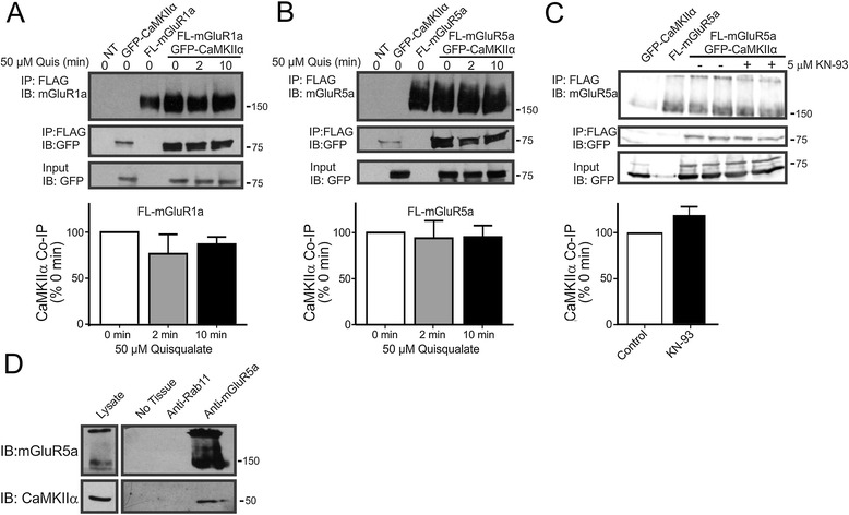
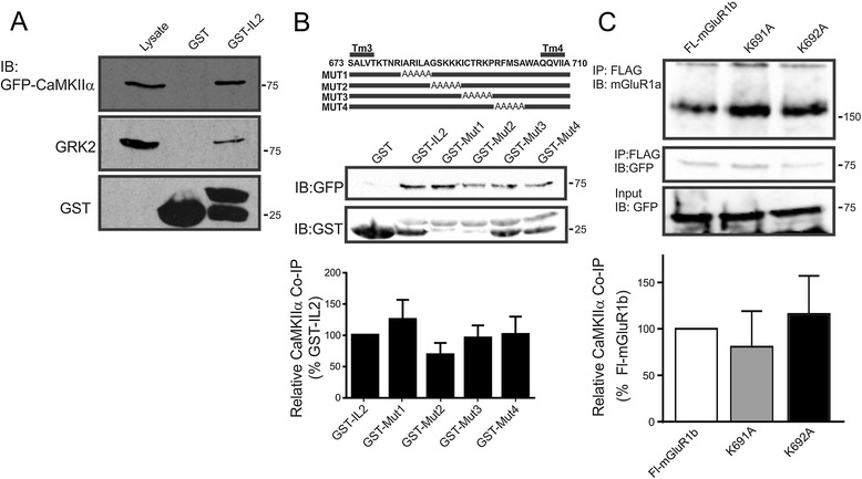
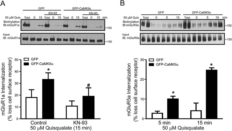
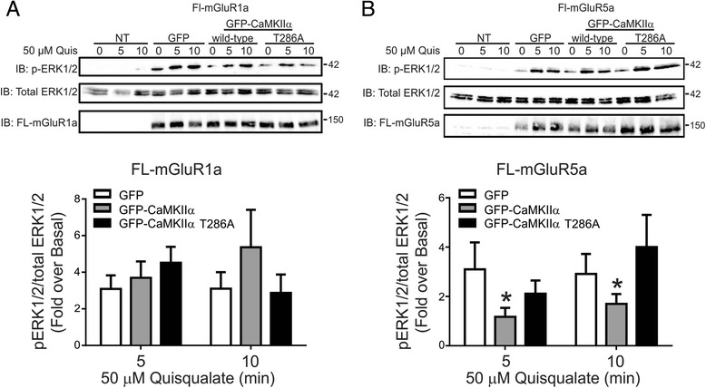
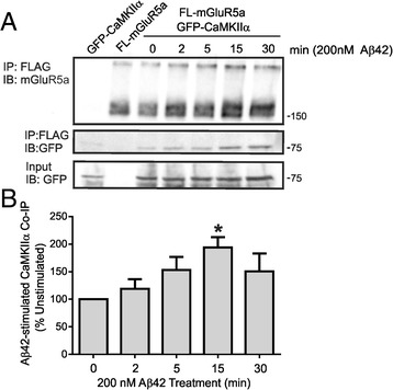
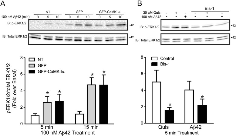
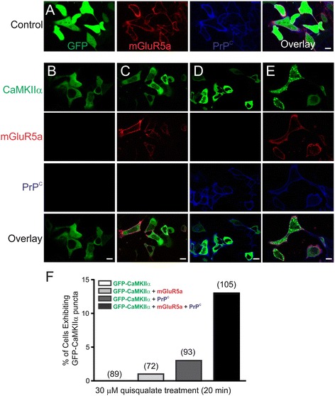
Results: In the present study, we utilized mass spectroscopy to identify calmodulin kinase IIα (CaMKIIα) as a protein that interacts with the second intracellular loop domain of mGluR5. We show that CaMKIIα interacts with both mGluR1a and mGluR5a in an agonist-independent manner and is co-immunoprecipitated with mGluR5a from hippocampal mouse brain. CaMKIIα positively regulates both mGluR1a and mGluR5a endocytosis, but selectively attenuates mGluR5a but not mGluR1a-stimulated ERK1/2 phosphorylation in a kinase activity-dependent manner. We also find that Aβ42 oligomers stimulate the association of CaMKIIα with mGluR5a and activate ERK1/2 in an mGluR5a-dependent manner. However, Aβ42 oligomer-stimulated ERK1/2 phosphorylation is not regulated by mGluR5a/CaMKIIα interactions suggesting that agonist and Aβ42 oligomers stabilize distinct mGluR5a activation states that are differentially regulated by CaMKIIα. The expression of both mGluR5a and PrP(C) together, but not alone resulted in the agonist-stimulated subcellular distribution of CaMKIIα into cytoplasmic puncta.

Conclusions: Taken together these results indicate that CaMKIIα selectively regulates mGluR1a and mGluR5a ERK1/2 signaling. As mGluR5 and CaMKIIα are involved in learning and memory and Aβ and mGluR5 are implicated in Alzheimer's disease, results of these studies could provide insight into potential pharmacological targets for treatment of Alzheimer's disease.


Figures

Figure 1
Figure 1
Co-immunoprecipitation of GFP-CaMKIIα with FLAG-mGluR1a and FLAG-mGluR5a. Representative immunoblot showing GFP-CaMKIIα co-immunoprecipitation with either A) FL-mGluR1a or B) FL-mGluR5a from HEK 293 cells transiently transfected as labeled with 3 μg of pcDNA3.1 of either FL-mGluR1a or FL-mGluR5a along with either 0.5 μg of plasmid cDNA encoding either pEGFP or GFP-CaMKIIα and treated with 50 μM quisqualate for the times indicated in the Figure. Bar graphs show the quantitative densitometric analysis of GFP-CaMKIIα co-immunoprecipitated with either FL-mGluR1a or FL-mGluR5a. The data represents the mean ± SD of 6 independent experiments. C) Representative immunoblot showing GFP-CaMKIIα co-immunoprecipitation with FLAG-mGluR5a in HEK 293 cells treated with or without 5 μM KN-93 that were transiently transfected with 2 μg of FL-mGluR5 along with 0.5 μg of GFP-CaMKIIα. Bar graphs show the quantitative densitometric analysis of GFP-CaMKIIα co-immunoprecipitated with FL-mGluR5a in the absence and following pretreatment with 5 μM KN-93 for 1 h. The data represents the mean ± SD of 6 independent experiments. D) Shown is a representative immunoblot of endogenous CaMKII co-immunoprecipitated with endogenous mGluR5. 1 mg of adult CD-1 mouse hippocampal tissue lysate was incubated with protein G sepharose beads and either polyclonal rabbit anti-Rab11 or anti-mGluR5 antibody. Shown is a representative immunoblot from 4 independent experiments.
Figure 2
Figure 2
Purified GST-fusion proteins encoding the mGluR1/5 IL2 loop domain interact with GFP-CaMKIIα. A) Shown is a representative immunoblot showing the co-immunoprecipitation of G protein-coupled receptor kinase 2 and GFP-CaMKIIα with a GST mGluR1/5 IL2 domain fusion protein. 1 μg of GST protein was incubated with 500 μg of HEK293 cell lysates over-expressing either GRK2 or GFP-CaMKIIα. The immunoblots are representative of 3 independent experiments. B) Upper panel, shows the schematic representation of GST-IL2 fusion protein alanine scanning mutants previously used to identify the IL2 residues required for GRK2 binding to Group I mGluRs [39]. Middle panel shows a representative immunoblot showing the co-immunoprecipitation of GFP-CaMKIIα with the GST-IL2 fusion protein alanine scanning mutants and the expression of the GST fusions. Lower panel shows the densitometric analysis of the relative co-immunoprecipitation of GFP-CaMKIIα with the GST-IL2 fusion protein alanine scanning mutants compared to the wild-type IL2 GST fusion protein. The data represents the mean ± SD of 3 independent experiments. C) Representative immunoblots showing GFP-CaMKIIα co-immunoprecipitated FL-mGluR1b and FL-mGluR1b mutants (K691A and K692A) that do not bind GRK2. Also shown are FL-mGluR1a immunoprecipitates and GFP-CaMKIIα expression in cell lysates. Bar graph shows the relative co-immunoprecipitation of GFP-CaMKIIα with the FL-mGluR1b and FL-mGluR1b mutants (K691A and K692A). Data shown represents the means ± SD of three independent experiments.
Figure 3
Figure 3
CaMKIIα overexpression increases agonist-stimulated mGluR1a and mGluR5 endocytosis. A) Upper blot shows a representative immunoblot for cell surface biotin-labeled mGluR1a in HEK 293 cells transfected with 3 μg of pcDNA3.1 encoding FL-mGluR1a along with 0.5 μg of plasmid cDNA encoding either GFP or GFP-CaMKIIα following 50 μM quisqualate treatment following 1 h pre-treatment in the presence and absence of KN-93. Lower blot shows the total cell lysates (50 μg) for mGluR1a. The bar graph shows the densitometric analysis of biotin-labeled cell surface mGluR1a immunoblots normalized to total mGluR1a biotinylation. Data represents the mean ± SD of 7 independent experiments. *P < 0.05 versus GFP transfected cells. B) Upper blot shows a representative immunoblot for cell surface biotin-labeled mGluR5a in HEK 293 cells transfected with 3 μg of pcDNA3.1 encoding FL-mGluR5a along with 0.5 μg of plasmid cDNA encoding either GFP or GFP-CaMKIIα following 50 μM quisqualate treatment. Lower blot shows the total cell lysates (50 μg) for mGluR5a. The bar graph shows the densitometric analysis of biotin-labeled cell surface mGluR1a immunoblots normalized to total mGluR5a biotinylation. Data represents the mean ± S.E.M. of 4 independent experiments completed in duplicate. *P < 0.05 versus GFP transfected cells, #P < 0.05 versus GFP CaMKIIα control.
Figure 4
Figure 4
Effect of CaMKIIα overexpression on mGluR1a- and mGluR5a- -stimulated ERK1/2 phosphorylation. A) Shown are representative immunoblots for FL-mGluR1a expression, p-ERK1/2 activity and total-ERK1/2 expression in HEK 293 cells transiently transfected with 3 μg of pcDNA3.1 encoding FLAG-mGluR1a along with 0.5 μg of plasmid cDNA encoding either GFP, GFP-CaMKIIα or GFP-CaMKIIα-T286A in response to 50 μM quisqualate treatment for 0, 5 and 10 min. Bar graph shows the densitometric analysis of ERK1/2 phosphorylation normalized to both basal and total ERK1/2 protein expression. Data represents the mean ± SD of five independent experiments. *P < 0.05 versus GFP transfected control cells. B) Shown are representative immunoblots for, FL-mGluR5a expression, p-ERK1/2 activity and total-ERK1/2 expression in HEK 293 cells transiently transfected with 3 μg of pcDNA3.1 encoding, FL-mGluR5a along with 0.5 μg of plasmid cDNA encoding either GFP, GFP-CaMKIIα or GFP-CaMKIIα-T286A in response to 50 μM quisqualate treatment for 0, 5 and 10 min. Bar graph shows the densitometric analysis of ERK1/2 phosphorylation normalized to both basal total ERK1/2 protein expression. Data represents the mean ± SD of five independent experiments. *P < 0.05 versus GFP transfected control cells.
Figure 5
Figure 5
Aβ42 oligomers increase CaMKIIα co-immunoprecipitation with mGluR5a. A) HEK 293 cells were transiently transfected with 2 μg of pcDNA3.1 encoding FLAG-mGluR5a and 0.5 μg of plasmid cDNA encoding GFP-CaMKIIα. HEK 293 cells were treated with 200 nM of Aβ42 oligomer for 0, 2, 5, 15 and 30 min. B) The bar graph shows the densitometric analysis of the relative co-immunoprecipitation of GFP-CaMKIIα with FL-mGluR5a following Aβ42 oligomer treatment normalized to total mGluR5a protein expression. Data represents the mean ± SD of 5 independent experiments. *p < 0.05 versus untreated cells.
Figure 6
Figure 6
Aβ42 oligomers stimulate ERK1/2 phosphorylation in an mGluR5a and PKC dependent manner that is independent of CaMKIIα overexpression. A) Shown are representative immunoblots of p-ERK1/2 activity and total ERK1/2 expression in HEK 293 cells transfected with 2 μg of pcDNA3.1 encoding FLAG-mGluR5a along with either 0.5 μg of plasmid cDNA encoding either GFP or CaMKIIα and treated with 100 nM Aβ42 oligomer for 5 and 10 min. Bar graph shows the densitometric analysis of ERK1/2 phosphorylation normalized to both basal and total ERK1/2 protein expression. Data represents the mean ± SD of 4 independent experiments. *P < 0.05 versus non-transfected (NT) cells. B) Shown are representative immunoblots of p-ERK1/2 activity and total ERK1/2 expression in HEK 293 cells transfected with 2 μg of pcDNA3.1 encoding FLAG-mGluR5a and treated with either 50 μM quisqualate or 100 nM Aβ42 oligomer in either the presence or absence of 1 μM Bis-1. Bar graph shows the densitometric analysis of ERK1/2 phosphorylation normalized to both basal and total ERK1/2 protein expression. Data represents the mean ± SD of 7 independent experiments. *P < 0.05 versus non-transfected (NT) cells.
Figure 7
Figure 7
Effect of mGluR5a and PrP C expression of GFP-CaMKIIα subcellular localization following agonist stimulation. HEK 293 cells were transiently transfected with different combinations of GFP-CaMKIIα (0.5 μg), FLAG-mGluR5a (2 μg), PrPC (2 μg) or empty pEGFP (2 μg) pcDNA3.1 plasmid cDNA. Shown are representative confocal microscopic images of HEK 293 cells transfected with A) pEGFP (control) along with both FL-mGluR5a and PrPC, B) GFP-CaMKIIα alone (green), C) GFP-CaMKIIα along with FL-mGluR5a (red), D) GFP-CaMKIIα along with PrPC (blue), and E) GFP-CaMKIIα along with both FL-mGluR5a and PrPC. All transfections were treated with 30 μM quisqualate for 20 min. F) Quantification of the number of cells exhibiting GFP-CaMKIIα puncta, number of cells imaged is shown in brackets. Data is representative 4 different experiments.

References

    1. Ferguson SSG. Evolving concepts in G protein-coupled receptor endocytosis: the role in receptor desensitization and signaling. Pharmacol Rev. 2001;53:1–24. - PubMed
    1. Walther C, Ferguson SSG. Role of arrestins in the desensitization, sequestration and vesicular trafficking of G protein-coupled receptors. Prog Mol Biol Transl Sci. 2013;118:93–113. doi: 10.1016/B978-0-12-394440-5.00004-8. - DOI - PubMed
    1. Dhami GK, Ferguson SSG. Metabotropic glutamate receptor desensitization. Pharmacol Ther. 2006;111:260–271. doi: 10.1016/j.pharmthera.2005.01.008. - DOI - PubMed
    1. Ferguson SSG. Phosphorylation-independent attenuation of GPCR signaling. Trends Pharmacol Sci. 2007;28:173–179. doi: 10.1016/j.tips.2007.02.008. - DOI - PubMed
    1. Ribeiro FM, Paquet M, Cregan SP, Ferguson SSG. Group I metabotropic glutamate receptor signaling and its implication in Neurological disease processes. CNS Neurol Disord: Drug Targets. 2010;9:574–595. doi: 10.2174/187152710793361612. - DOI - PubMed

Publication types

MeSH terms

Substances