

Immunoproteomic identification of immunodominant antigens independent of the time of infection in Brucella abortus 2308-challenged cattle
- PMID: 25885057
- PMCID: PMC4345015
- DOI: 10.1186/s13567-015-0147-6
Immunoproteomic identification of immunodominant antigens independent of the time of infection in Brucella abortus 2308-challenged cattle
Abstract
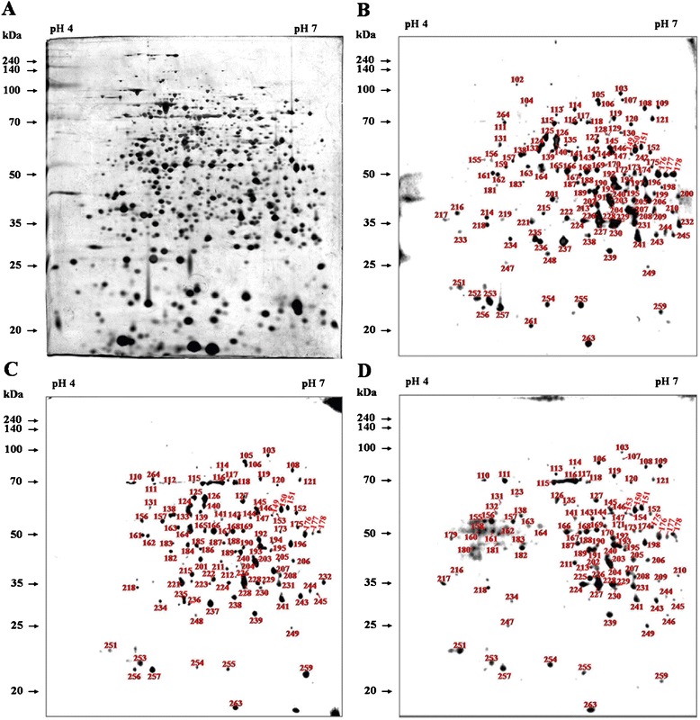
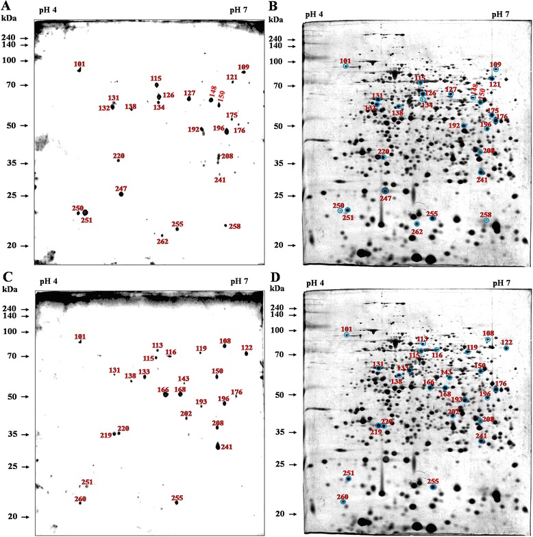
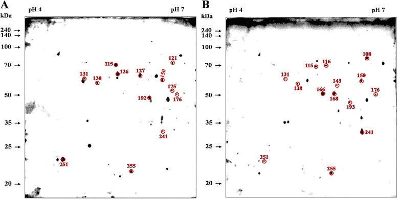
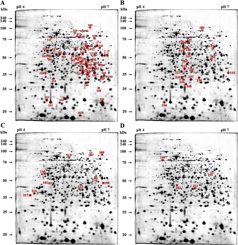
Brucellosis is a vital zoonotic disease caused by Brucella, which infects a wide range of animals and humans. Accurate diagnosis and reliable vaccination can control brucellosis in domestic animals. This study examined novel immunogenic proteins that can be used to detect Brucella abortus infection or as an effective subcellular vaccine. In an immunoproteomic assay, 55 immunodominant proteins from B. abortus 544 were observed using two dimensional electrophoresis (2DE) and immunoblot profiles with antisera from B. abortus-infected cattle at the early (week 3), middle (week 7), and late (week 10) periods, after excluding protein spots reacting with antisera from Yersinia enterocolitica O:9-infected and non-infected cattle. Twenty-three selected immunodominant proteins whose spots were observed at all three infection periods were identified using MALDI-MS/MS. Most of these proteins identified by immunoblot and mass spectrometry were determined by their subcellular localization and predicted function. We suggest that the detection of prominent immunogenic proteins during the infection period can support the development of advanced diagnostic methods with high specificity and accuracy; subsidiarily, these proteins can provide supporting data to aid in developing novel vaccine candidates.
Figures
Similar articles
-
Proteomic analyses of the time course responses of mice infected with Brucella abortus 544 reveal immunogenic antigens.FEMS Microbiol Lett. 2014 Aug;357(2):164-74. doi: 10.1111/1574-6968.12522. Epub 2014 Jul 22. FEMS Microbiol Lett. 2014. PMID: 24975114
-
Comprehensive Identification of Immunodominant Proteins of Brucella abortus and Brucella melitensis Using Antibodies in the Sera from Naturally Infected Hosts.Int J Mol Sci. 2016 Apr 30;17(5):659. doi: 10.3390/ijms17050659. Int J Mol Sci. 2016. PMID: 27144565 Free PMC article.
-
Immunogenic proteins of Brucella abortus to minimize cross reactions in brucellosis diagnosis.Vet Microbiol. 2012 May 4;156(3-4):374-80. doi: 10.1016/j.vetmic.2011.11.011. Epub 2011 Nov 22. Vet Microbiol. 2012. PMID: 22192360
-
Molecular and cellular interactions between Brucella abortus antigens and host immune responses.Vet Microbiol. 2002 Dec 20;90(1-4):417-24. doi: 10.1016/s0378-1135(02)00225-0. Vet Microbiol. 2002. PMID: 12414160 Review.
-
Immune response triggered by Brucella abortus following infection or vaccination.Vaccine. 2015 Jul 17;33(31):3659-66. doi: 10.1016/j.vaccine.2015.05.057. Epub 2015 Jun 3. Vaccine. 2015. PMID: 26048781 Review.
Cited by
-
Heat-stress-modulated induction of NF-κB leads to brucellacidal pro-inflammatory defense against Brucella abortus infection in murine macrophages and in a mouse model.BMC Microbiol. 2018 May 25;18(1):44. doi: 10.1186/s12866-018-1185-9. BMC Microbiol. 2018. PMID: 29801438 Free PMC article.
-
Proteomics of Brucella: Technologies and Their Applications for Basic Research and Medical Microbiology.Microorganisms. 2020 May 20;8(5):766. doi: 10.3390/microorganisms8050766. Microorganisms. 2020. PMID: 32443785 Free PMC article.
-
Expression of cytokine and apoptosis-related genes in bovine peripheral blood mononuclear cells stimulated with Brucella abortus recombinant proteins.Vet Res. 2016 Feb 11;47:30. doi: 10.1186/s13567-016-0311-7. Vet Res. 2016. PMID: 26864657 Free PMC article.
-
Loci Associated With Antibody Response in Feral Swine (Sus scrofa) Infected With Brucella suis.Front Vet Sci. 2020 Nov 25;7:554674. doi: 10.3389/fvets.2020.554674. eCollection 2020. Front Vet Sci. 2020. PMID: 33324693 Free PMC article.
-
Comparative evaluation of the immunodominant proteins of Brucella abortus for the diagnosis of cattle brucellosis.Vet World. 2021 Mar;14(3):803-812. doi: 10.14202/vetworld.2021.803-812. Epub 2021 Mar 30. Vet World. 2021. PMID: 33935431 Free PMC article.
References
-
- McGiven JA. New developments in the immunodiagnosis of brucellosis in livestock and wildlife. Rev Sci Tech. 2013;32:163–176. - PubMed
-
- Nielsen K, Yu WL. Serological diagnosis of brucellosis. Prilozi. 2010;31:65–89. - PubMed
-
- McGiven JA, Stack JA, Perrett LL, Tucker JD, Brew SD, Stubberfield E, MacMillan AP. Harmonisation of European tests for serological diagnosis of Brucella infection in bovines. Rev Sci Tech. 2006;25:1039–1053. - PubMed
Publication types
MeSH terms
Substances
LinkOut - more resources
Full Text Sources
Other Literature Sources
