. 2015 Apr 17;10(4):e0124802.
doi: 10.1371/journal.pone.0124802. eCollection 2015.

Interferon-β suppresses murine Th1 cell function in the absence of antigen-presenting cells

Affiliations

Interferon-β suppresses murine Th1 cell function in the absence of antigen-presenting cells

Nicolas Boivin et al. PLoS One. .

Abstract

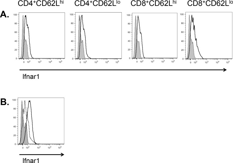
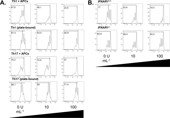
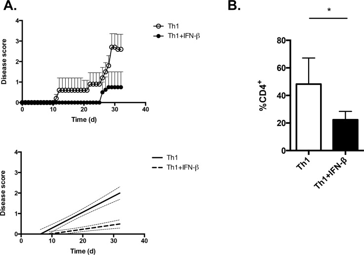
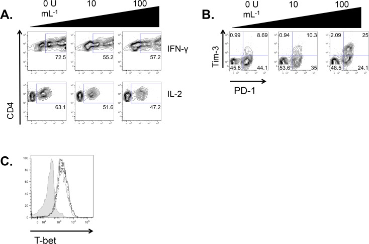
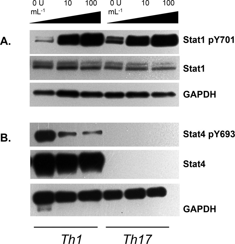
Interferon (IFN)-β is a front-line therapy for the treatment of the relapsing-remitting form of multiple sclerosis. However, its immunosuppressive mechanism of function remains incompletely understood. While it has been proposed that IFN-β suppresses the function of inflammatory myelin antigen-reactive T cells by promoting the release of immunomodulatory cytokines such as IL-27 from antigen-presenting cells (APCs), its direct effects on inflammatory CD4+ Th1 cells are less clear. Here, we establish that IFN-β inhibits mouse IFN-γ+ Th1 cell function in the absence of APCs. CD4+ T cells express the type I interferon receptor, and IFN-β can suppress Th1 cell proliferation under APC-free stimulation conditions. IFN-β-treated myelin antigen-specific Th1 cells are impaired in their ability to induce severe experimental autoimmune encephalomyelitis (EAE) upon transfer to lymphocyte-deficient Rag1-/- mice. Polarized Th1 cells downregulate IFN-γ and IL-2, and upregulate the negative regulatory receptor Tim-3, when treated with IFN-β in the absence of APCs. Further, IFN-β treatment of Th1 cells upregulates phosphorylation of Stat1, and downregulates phosphorylation of Stat4. Our data indicate that IFN-γ-producing Th1 cells are directly responsive to IFN-β and point to a novel mechanism of IFN-β-mediated T cell suppression that is independent of APC-derived signals.


Conflict of interest statement

Competing Interests: The work was supported by funds from the CHU de Québec, and by a EMD Serono, Canada and end MS Research and Training Network Transitional Career Development Award from the MS Society of Canada and the Multiple Sclerosis Scientific Research Foundation. Acceptance of this unrestricted educational grant by the MS Society of Canada does not constitute endorsement by researcher or the Society of any product(s) of EMD Serono Canada Company. The MS Society of Canada does not approve, endorse or recommend any specific product or therapy but provides information to assist individuals in making their own decisions. The MS Society of Canada is an independent, voluntary health agency and does not approve, endorse or recommend any specific product or therapy but provides information to assist individuals in making their own decisions. Acceptance of this award does not alter the authors’ adherence to PLOS ONE policies on sharing data and materials.

Figures

Fig 1
Fig 1. Th1 cells express the type I interferon receptor.
A. Naïve (CD62Lhi) and effector (CD62Llo) CD4+ and CD8+ C57BL/6J T cells were assessed for surface expression of Ifnar1 by flow cytometry. Open histogram, Ifnar1; shaded histogram, fluorescence minus one (FMO) control. B. Th1 and Th17 cells were cultured and assessed for surface expression of Ifnar1 after 5 days. Solid line with open histogram, Th1; Dotted line with open histogram, Th17; shaded histogram, FMO control. Data representative of one of three experiments.
Fig 2
Fig 2. IFN-β suppresses Th1 cell proliferation.
A. WT CD4+CD62Lhi T cells were labeled with CFSE and stimulated for 5 days with either soluble anti-CD3 plus irradiated splenocytes (APCs), or with plate-bound anti-CD3+anti-CD28, under Th1 or Th17 conditions, with the indicated concentrations of IFN-β. CFSE dilution was assessed by flow cytometry. Data representative of three experiments. B. WT or IFNAR1-/- CD4+CD62Lhi T cells were labeled with CFSE and stimulated with plate-bound anti-CD3+anti-CD28 under Th1 conditions with the indicated concentrations of IFN-β for 5 days. CFSE dilution was assessed by flow cytometry. Data representative of two experiments. Gates represent the percentage of cells that underwent at least one division.
Fig 3
Fig 3. IFN-β suppresses the encephalitogenic potential of Th1 cells.
CD4+CD62Lhi T cells were isolated from female 2D2 mouse spleens and lymph nodes, and were stimulated with plate-bound anti-CD3+anti-CD28 under Th1 conditions, in the presence or absence of 100 U mL-1 of IFN-β, for 5 days. They were then transferred to female, 6-week old, Rag1-/- mice (5x10⁶ cells/mouse). Recipient mice were monitored for clinical signs of EAE. n = 5 Th1, n = 4 Th1+IFN-β. Right graph, linear regression curves of the disease courses. The slopes are significantly different between the disease courses (p<0.0006). B. Brains and spinal cords were isolated from mice in (A) at d32 and single mononuclear cell suspensions were obtained. The frequency of CNS-infiltrating CD4+ cells was assessed by flow cytometry. * p<0.05, two-tailed Student’s t test.
Fig 4
Fig 4. IFN-β suppresses cytokine secretion from Th1 cells.
A. CD4+CD62Lhi T cells were sorted from the spleens and lymph nodes of C57BL6/J mice and were stimulated with plate-bound anti-CD3+anti-CD28 under Th1 conditions, with the indicated concentrations of IFN-β, for 5 days. Cells were restimulated for 48 hours under Th1 conditions with the same concentration of IFN-β as at the initial stimulation. A. Generation of IFN-γ and IL-2 was assessed by intracellular cytokine staining and flow cytometry. Representative of three experiments. B. Expression of Tim-3 and PD-1 were assessed by flow cytometry. Representative of three experiments. C. Expression of T-bet was assessed by flow cytometry. Representative data from one of three mice assessed individually. Solid line with open histogram, Th1; dashed line with open histogram, Th1 + 10 U mL-1 IFN-β; dotted line with open histogram, Th1 + 100 U mL-1 IFN-β; shaded histogram, FMO control. Data in all panels gated on CD4+ cells.
Fig 5
Fig 5. IFN-β regulates Stat1 and Stat4 expression on Th cells in the absence of APCs.
CD4+CD62LhiCD25- T cells were sorted from the spleens and lymph nodes of C57BL6/J mice. They were stimulated with plate-bound anti-CD3+anti-CD28 under Th1 or Th17 conditions, with the indicated concentrations of IFN-β, for 48 hours, and cell lysates were generated. A. Expression of Stat1 and pStat1(Y701) were assessed by Western blot. Representative of two experiments. B. Expression of Stat4 and pStat4(Y693) were assessed by Western blot. Representative of three experiments. GAPDH, loading control.

References

    1. Beck CA, Metz LM, Svenson LW, Patten SB. Regional variation of multiple sclerosis prevalence in Canada. Mult Scler. 2005; 11: 516–519. - PubMed
    1. Ascherio A. Environmental factors in multiple sclerosis. Expert Rev Neurother. 2013; 13: 3–9. 10.1586/14737175.2013.865866 - DOI - PubMed
    1. Nyland H, Mörk S, Matre R. In-situ characterization of mononuclear cell infiltrates in lesions of multiple sclerosis. Neuropathol Appl Neurobiol. 1982; 8: 403–411. - PubMed
    1. International Multiple Sclerosis Genetics Consortium, Wellcome Trust Case Control Consortium 2, Sawcer S, Hellenthal G, Pirinen M, Spencer CC, Patsopoulos NS, et al. Genetic risk and a primary role for cell-mediated immune mechanisms in multiple sclerosis. Nature. 2011; 476: 214–219. 10.1038/nature10251 - DOI - PMC - PubMed
    1. Weber MS, Hohlfeld R, Zamvil SS. Mechanism of action of glatiramer acetate in treatment of multiple sclerosis. Neurotherapeutics. 2007; 4: 647–653. 10.1016/j.nurt.2007.08.002 - DOI - PMC - PubMed

Publication types

Substances