. 2015 Feb 22;16(1):42.
doi: 10.1186/s13059-015-0594-4.

Methylation-dependent SOX9 expression mediates invasion in human melanoma cells and is a negative prognostic factor in advanced melanoma

Affiliations

Methylation-dependent SOX9 expression mediates invasion in human melanoma cells and is a negative prognostic factor in advanced melanoma

Phil F Cheng et al. Genome Biol. .

Abstract

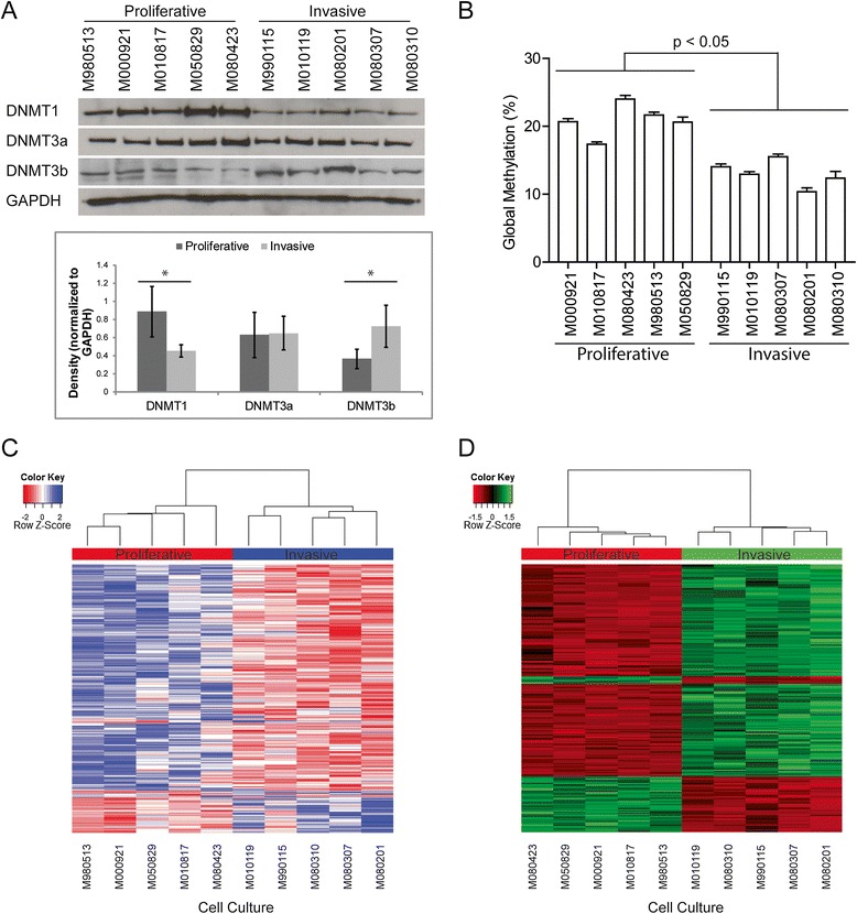
Background: Melanoma is the most fatal skin cancer displaying a high degree of molecular heterogeneity. Phenotype switching is a mechanism that contributes to melanoma heterogeneity by altering transcription profiles for the transition between states of proliferation/differentiation and invasion/stemness. As phenotype switching is reversible, epigenetic mechanisms, like DNA methylation, could contribute to the changes in gene expression.

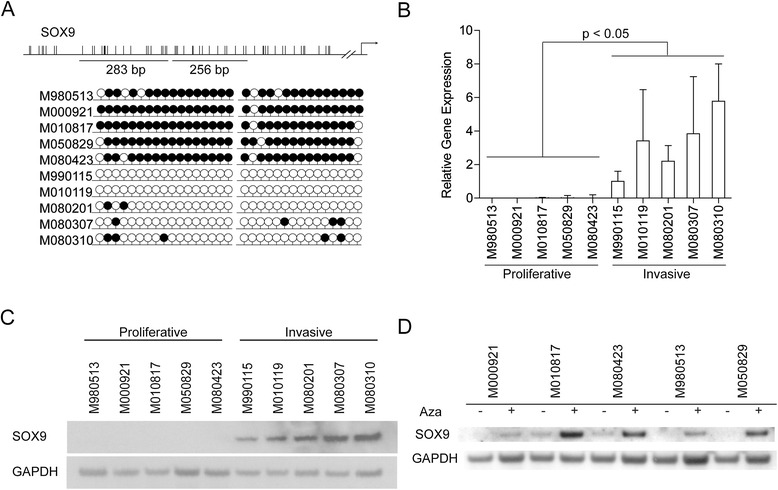
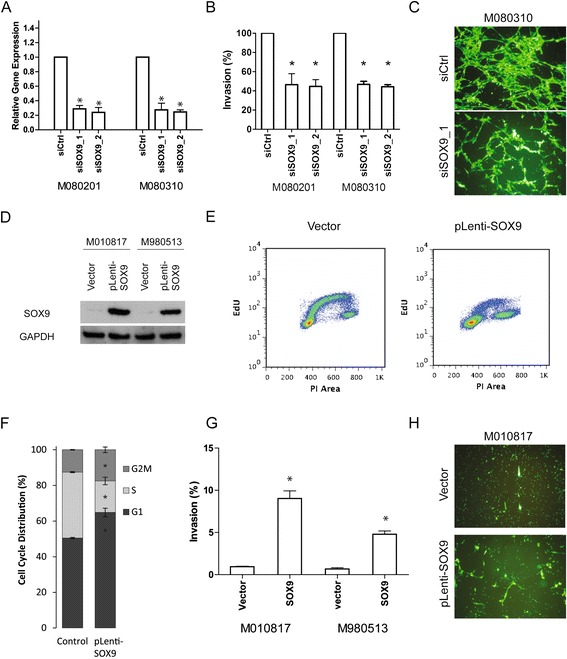
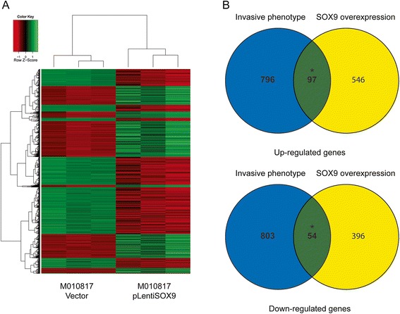
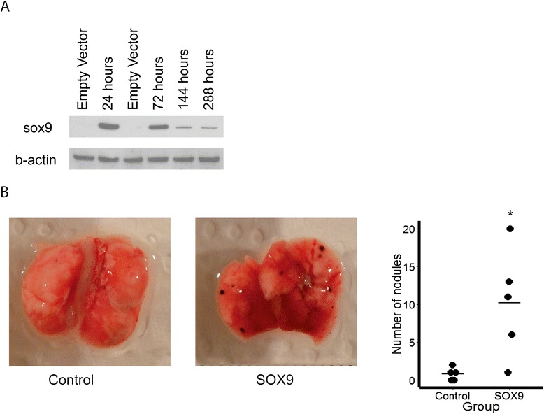
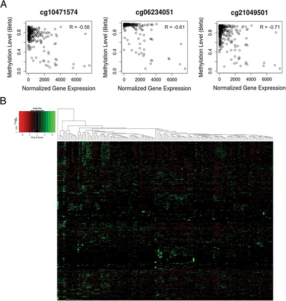
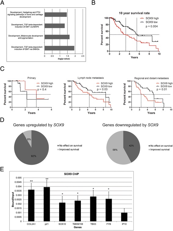
Results: Integrative analysis of methylation and gene expression datasets of five proliferative and five invasion melanoma cell cultures reveal two distinct clusters. SOX9 is methylated and lowly expressed in the highly proliferative group. SOX9 overexpression results in decreased proliferation but increased invasion in vitro. In a B16 mouse model, sox9 overexpression increases the number of lung metastases. Transcriptional analysis of SOX9-overexpressing melanoma cells reveals enrichment in epithelial to mesenchymal transition (EMT) pathways. Survival analysis of The Cancer Genome Atlas melanoma dataset shows that metastatic patients with high expression levels of SOX9 have significantly worse survival rates. Additional survival analysis on the targets of SOX9 reveals that most SOX9 downregulated genes have survival benefit for metastatic patients.

Conclusions: Our genome-wide DNA methylation and gene expression study of 10 early passage melanoma cell cultures reveals two phenotypically distinct groups. One of the genes regulated by DNA methylation between the two groups is SOX9. SOX9 induces melanoma cell invasion and metastasis and decreases patient survival. A number of genes downregulated by SOX9 have a negative impact on patient survival. In conclusion, SOX9 is an important gene involved in melanoma invasion and negatively impacts melanoma patient survival.


Figures

Figure 1
Figure 1
Phenotype specific expression of DNMTs and DNA methylation patterns. (A) Western blot for DNMT1 and DNMT3a and DNMT3b on five proliferative phenotype and five invasive phenotype cell lysates. GAPDH was used as a loading control. Optical density of each band was measured and normalized to GAPDH intensity. (B) Global methylation analysis by ELISA for methyl-cytosine on five proliferative cell cultures and five invasive cell cultures. There was a significant difference (P <0.05, Student’s t-test) in global methylation between the proliferative and invasive phenotype melanoma cells. (C) Heat map representing the 406 gene promoters differentially methylated between the proliferative and invasive phenotype cultures. (D) Heat map representing the top 250 genes differentially expressed between the proliferative and invasive phenotype.
Figure 2
Figure 2
Validation of SOX9 methylation. (A) Lollipop diagrams of bisulphite sequencing of SOX9 promoter. Black lollipops are methylated CpGs; white lollipops are unmethylated CpGs. A minimum of five clones were sequenced for each cell culture. (B) mRNA expression of SOX9 normalized to the housekeeping gene RPL28 across 10 melanoma cell cultures. Results are presented as mean +/− s.d., n = 3. Statistical significance of differential expression between the proliferative and invasive phenotype cell cultures was determined by Student’s t-test. (C) Western blot for SOX9 in 10 melanoma cell cultures. GAPDH served as loading control. (D) Western blot for SOX9 of five proliferative melanoma cell cultures treated with 5-aza-2-deoxycytidine (Aza).
Figure 3
Figure 3
SOX9 mediates invasion and cell cycle arrest. (A) SOX9 knockdown by siRNA in two melanoma cell cultures M080201 and M080310. (B) Boyden chamber assay for siSOX9 knockdown in M080201 and M080310. (C) Representative picture of the Boyden chamber assay for knockdown of SOX9 in M080310. Top panel shows M080310 cells treated with control siRNA (siCtrl). Bottom panel shows M080310 cells treated with siSOX9_1. (D) SOX9 overexpression by lentiviral expression in proliferative phenotype melanoma cell cultures M010817 and M980513. (E) Proliferation assay by EdU pulse in cell cultures transfected with empty vector and pLenti-SOX9. (F) Cell cycle analysis by PI staining for cell cultures transfected with empty vector and pLenti-SOX9 (top and bottom left panels). Quantitation of cell cycle analysis (* = P <0.05, right panel). (G) Boyden chamber assay for SOX9 overexpressing melanoma cell cultures M010817 and M980513 (* = P <0.05). (H) Representative picture of the Boyden chamber assay for SOX9 overexpression in M010817. Top panel shows M010817 transfected with empty vector. Bottom panel shows M010817 transfected with SOX9.
Figure 4
Figure 4
Microarray analysis of SOX9 overexpression. (A) Heat map of M010817 cells overexpressing SOX9. (B) Overlap of upregulated genes and downregulated genes between the SOX9 microarray and 10 melanoma cell culture microarray.
Figure 5
Figure 5
In vivo function of sox9 overexpression. (A) Western blot analysis of transient sox9 overexpression in B16F1 up to 12 days. (B) Representative pictures of B16F1 cells intravenously injected into C57BL/6 J mice 12 days after injection (n = 5 mice per group). Quantification (right graph). * = P <0.05 Mann–Whitney U test.
Figure 6
Figure 6
Validation of SOX9 methylation in TCGA and correlation with clinical features. (A) Correlation plots of RNAseq reads to b-values of methylation for SOX9. Three methylation probes of SOX9 are shown here to have significant anti-correlation of gene expression and DNA methylation. (B) Heatmap of the 427 genes differentially expressed between the SOX9 high and SOX9 low metastatic melanoma samples.
Figure 7
Figure 7
TCGA analysis of SOX9. (A) Pathway analysis from MetaCore for SOX9 reveals many pathways involved in EMT. (B) Median survival time for patients with high SOX9 expression is 3.9 years (n = 74). Median survival time for patients with low SOX9 expression is 5.8 years (n = 74). The difference in survival is significant (P <0.05). (C) Survival analysis for high and low SOX9 expression segregated into primary, lymph node, and metastatic cohorts. (D) Pie charts displaying the number of SOX9 target genes that have a contribution to patient survival. (E) ChIP analysis for SOX9 binding targets. Enrichment of the promoter regions for TMEM158, TBX3, and FYB were similar to positive controls COL2A1, p21, and SOX10 and greater than negative control IP10. Data are shown as bound vs. input. Error bars indicate standard error of the mean of three independent experiments. IgG controls not shown on graph because they were below detection limit.

References

    1. Dupin E, Le Douarin NM. Development of melanocyte precursors from the vertebrate neural crest. Oncogene. 2003;22:3016–23. doi: 10.1038/sj.onc.1206460. - DOI - PubMed
    1. Garbe C, Leiter U. Melanoma epidemiology and trends. Clin Dermatol. 2009;27:3–9. doi: 10.1016/j.clindermatol.2008.09.001. - DOI - PubMed
    1. MacKie RM, Hauschild A, Eggermont AM. Epidemiology of invasive cutaneous melanoma. Ann Oncol. 2009;20:vi1–7. doi: 10.1093/annonc/mdp252. - DOI - PMC - PubMed
    1. Quintana E, Shackleton M, Foster HR, Fullen DR, Sabel MS, Johnson TM, et al. Phenotypic heterogeneity among tumorigenic melanoma cells from patients that is reversible and not hierarchically organized. Cancer Cell. 2010;18:510–23. doi: 10.1016/j.ccr.2010.10.012. - DOI - PMC - PubMed
    1. Perego M, Tortoreto M, Tragni G, Mariani L, Deho P, Carbone A, et al. Heterogeneous phenotype of human melanoma cells with in vitro and in vivo features of tumor-initiating cells. J Invest Dermatol. 2010;130:1877–86. doi: 10.1038/jid.2010.69. - DOI - PubMed

Publication types

MeSH terms

Substances