. 2015 Oct;9(10):2261-74.
doi: 10.1038/ismej.2015.39. Epub 2015 Apr 17.

The coral core microbiome identifies rare bacterial taxa as ubiquitous endosymbionts

Affiliations

The coral core microbiome identifies rare bacterial taxa as ubiquitous endosymbionts

Tracy D Ainsworth et al. ISME J. 2015 Oct.

Abstract

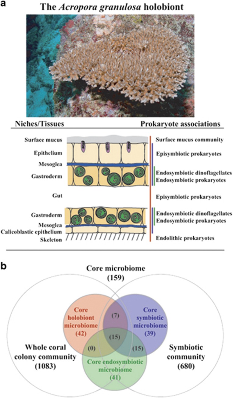
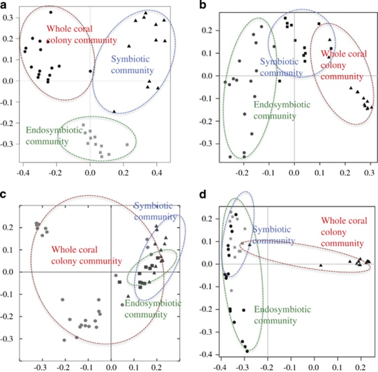
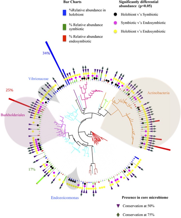
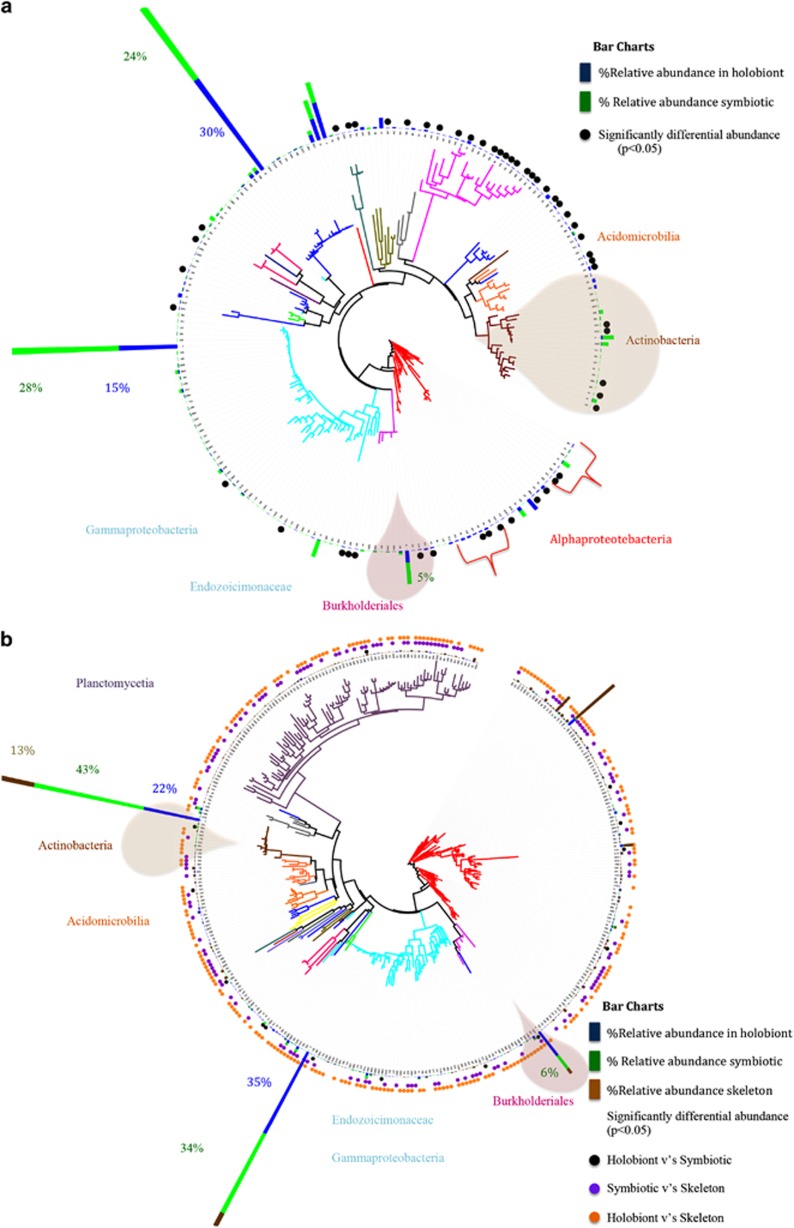
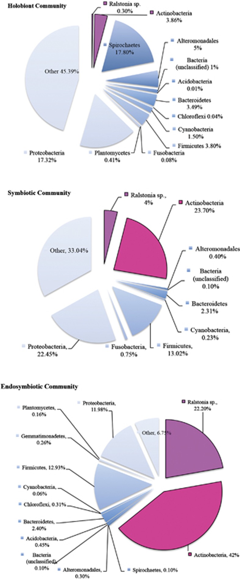
Despite being one of the simplest metazoans, corals harbor some of the most highly diverse and abundant microbial communities. Differentiating core, symbiotic bacteria from this diverse host-associated consortium is essential for characterizing the functional contributions of bacteria but has not been possible yet. Here we characterize the coral core microbiome and demonstrate clear phylogenetic and functional divisions between the micro-scale, niche habitats within the coral host. In doing so, we discover seven distinct bacterial phylotypes that are universal to the core microbiome of coral species, separated by thousands of kilometres of oceans. The two most abundant phylotypes are co-localized specifically with the corals' endosymbiotic algae and symbiont-containing host cells. These bacterial symbioses likely facilitate the success of the dinoflagellate endosymbiosis with corals in diverse environmental regimes.


Figures

Figure 1
Figure 1
The host coral Acropora granulosa (pictured), the tissues layers of the host coral and regions of bacterial association (a) and Venn diagram of the inclusion of niche habitat associations within the coral microbiome (b). Coral holobiont is indicated in red, coral symbiotic tissue community is indicated in blue and coral endosymbiotic community is indicated in green.
Figure 2
Figure 2
PCoA (Bray–Curtis) plot of bacterial associations within coral host habitats of the coral Acropora granulosa (a). PCoA (Bray–Curtis) plot of predicted metagenomes within the whole coral colony community (b). PCoA (Bray–Curtis) plot of bacterial associations within coral host habitats of the core microbiome of the coral A. granulosa (c). PCoA (Bray–Curtis) plot of predicted metagenomes within the microbiome of the coral A. granulosa (d).
Figure 3
Figure 3
Dendrogram (Tree of Life) of coral-associated bacteria demonstrating average relative abundance of coral-associated bacterial communities within specific tissue regions of Acropora granulosa from the Great Barrier Reef and Coral Sea. Blue abundance bars represent holobiont (whole community) core microbiome, green abundance bars represent the specific members of the endosymbiotic and episymbiotic core microbiome of the coral tissues (tissue associated) and red abundance bars represent the specific members of the core microbiome from endosymbiotic regions. Individual bacterial phylotypes found to have significantly differential relative abundance (P<0.05) between the holobiont and symbiotic community are shown in black, holobiont and endosymbiotic community are shown in yellow and symbiotic and endosymbtioic community are shown in pink. Bacterial classes are shown by color. 50% core annotation is represented by purple triangle; 75% core annotation is represented by green diamond.
Figure 4
Figure 4
Dendrogram (Tree of Life) of coral-associated bacteria demonstrating average relative abundance of coral-associated bacterial communities within specific tissue regions of Montipora capitata (a) and Leptoseris spp. (b) from Hawai‘i. Blue abundance bars represent core holobiont (whole community) microbiome, green abundance bars represent the specific members within the endosymbiotic and episymbiotic core microbiome of the coral tissues and brown bars represent the specific member within the core microbiome of skeleton-associated (endolithic) microbiome. Individual bacterial phylotypes found to have significantly different relative abundance between the holobiont and symbiotic community are shown in black, holobiont and skeletal community are shown in brown and symbiotic and skeletal community are shown in purple. Bacterial classes are shown by color.
Figure 5
Figure 5
The percentage of relative abundance of groupings within endosymbiotic core microbiome, endosymbiotic and episymbiotic core microbiome and holobiont core microbiome.
Figure 6
Figure 6
Localization of Actinobacter (ae) and Ralstonia sp. (fk), within the endosymbiotic dinoflagellates and gastrodermal cells of the coral host using FISH. Red indicates binding of the fluorescently labeled probe to bacteria cells within the host and endosymbiotic cells. Green indicates endosymbiotic dinoflagellates, blue indicates coral host tissues. Scale bar=10 μm.

References

    1. Ainsworth TD, Fine M, Blackall LL, Hoegh-Guldberg O. (2006). Fluorescence in situ hybridization and spectral imaging of coral-associated bacterial communities. Appl Environ Microbiol 72: 3016–3020. - PMC - PubMed
    1. Ainsworth TD, Thurber RV, Gates RD. (2010). The future of coral reefs: a microbial perspective. Trends Ecol Evol 25: 233–240. - PubMed
    1. Bäckhed F, Fraser Claire M, Ringel Y, Sanders Mary E, Sartor RB, Sherman Philip M et al. (2012). Defining a healthy human gut microbiome: current concepts, future directions, and clinical applications. Cell Host Microbe 12: 611–622. - PubMed
    1. Bayer T, Neave MJ, Alsheikh-Hussain A, Aranda M, Yum LK, Mincer T et al. (2013). The microbiome of the Red Sea coral Stylophora pistillata is dominated by tissue-associated Endozoicomonas bacteria. Appl Environ Microbiol 79: 4759–4762. - PMC - PubMed
    1. Bosch TCG, McFall-Ngai MJ. (2011). Metaorganisms as the new frontier. Zoology 114: 185–190. - PMC - PubMed

Publication types