. 2015 Apr 12:10:44.
doi: 10.1186/s13023-015-0256-3.

Phenotypic and molecular insights into CASK-related disorders in males

Affiliations

Phenotypic and molecular insights into CASK-related disorders in males

Ute Moog et al. Orphanet J Rare Dis. .

Abstract

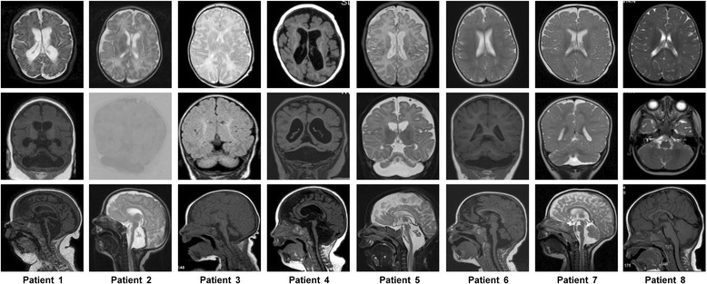
Background: Heterozygous loss-of-function mutations in the X-linked CASK gene cause progressive microcephaly with pontine and cerebellar hypoplasia (MICPCH) and severe intellectual disability (ID) in females. Different CASK mutations have also been reported in males. The associated phenotypes range from nonsyndromic ID to Ohtahara syndrome with cerebellar hypoplasia. However, the phenotypic spectrum in males has not been systematically evaluated to date.

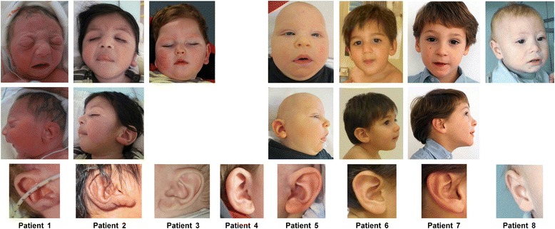
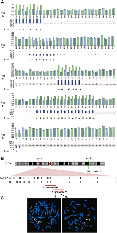
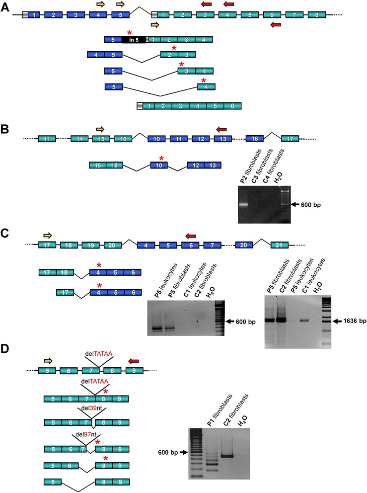
Methods: We identified a CASK alteration in 8 novel unrelated male patients by targeted Sanger sequencing, copy number analysis (MLPA and/or FISH) and array CGH. CASK transcripts were investigated by RT-PCR followed by sequencing. Immunoblotting was used to detect CASK protein in patient-derived cells. The clinical phenotype and natural history of the 8 patients and 28 CASK-mutation positive males reported previously were reviewed and correlated with available molecular data.

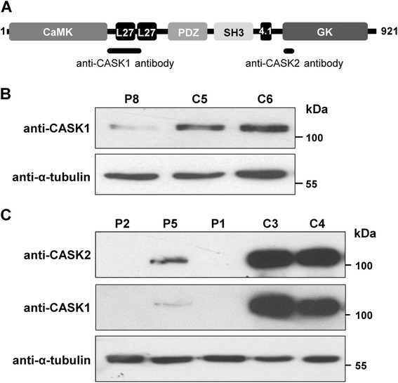
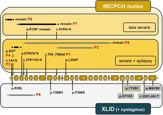
Results: CASK alterations include one nonsense mutation, one 5-bp deletion, one mutation of the start codon, and five partial gene deletions and duplications; seven were de novo, including three somatic mosaicisms, and one was familial. In three subjects, specific mRNA junction fragments indicated in tandem duplication of CASK exons disrupting the integrity of the gene. The 5-bp deletion resulted in multiple aberrant CASK mRNAs. In fibroblasts from patients with a CASK loss-of-function mutation, no CASK protein could be detected. Individuals who are mosaic for a severe CASK mutation or carry a hypomorphic mutation still showed detectable amount of protein.

Conclusions: Based on eight novel patients and all CASK-mutation positive males reported previously three phenotypic groups can be distinguished that represent a clinical continuum: (i) MICPCH with severe epileptic encephalopathy caused by hemizygous loss-of-function mutations, (ii) MICPCH associated with inactivating alterations in the mosaic state or a partly penetrant mutation, and (iii) syndromic/nonsyndromic mild to severe ID with or without nystagmus caused by CASK missense and splice mutations that leave the CASK protein intact but likely alter its function or reduce the amount of normal protein. Our findings facilitate focused testing of the CASK gene and interpreting sequence variants identified by next-generation sequencing in cases with a phenotype resembling either of the three groups.


Figures

Figure 1
Figure 1
Photographs of patients 1–8. Facial features of patients 1–8 are shown at the age of 1 day (patient 1), 9 months (patient 2, ear at 3 days), 19 months (patient 3), 7 months (patient 5), 16 months (patient 6), 5 years (patient 7), 14 months (patient 8), ears are depicted in the bottom row. Apart from microcephaly, patients 5 and 6 show epicanthal folds and a long philtrum, patient 8 a flat smooth philtrum, patients 1, 5 and 6 a broad and patient 7 a prominent nasal bridge, patient 1 a bulbous tip of nose, patients 1 and 5 retromicrognathia, and patients 1, 2, 3 and 5 fleshy, uplifted ear lobules. There seems to be no recognizable facial phenotype.
Figure 2
Figure 2
Selected axial, coronal and sagittal MR images from eight male individuals with a CASK alteration. The coronal images (second row) show hypoplastic, flattened cerebellar hemispheres with proportionally reduced size of the vermis in patients 1–7. The sagittal images in the third row show intact corpus callosum in all cases, low forehead indicative for microcephaly and pontine hypoplasia in patients 1–7. Pontocerebellar hypoplasia is severe in patients 1–5, moderate in patient 6, and mild in patient 7. Cerebellum and pons of patient 8 are normal. A mildly reduced number and complexity of the frontal gyri are seen in patients 1 and 7, and cortical atrophy in patients 1 and 4 (axial images in first row). MR imaging was performed at the age of 6 months (patient 1), 5 months (patient 2), 5 years (patient 3), 2 months (patient 4), 4 months (patient 5), 11 months (patient 6), 10 months (patient 7), and 16 months (patient 8).
Figure 3
Figure 3
Partial CASK deletions and duplications. A. MLPA results on DNA isolated from leukocytes of patients 2, 5, 6, and 7 and on lymphoblastoid cell-derived DNA of patient 8. Bars in upper and lower histograms represent MLPA probes. Peak area histogram (upper histogram): blue bars represent the mean probe signals with standard deviations for three reference DNAs, except for patient 7 (one reference DNA); green bars: probe signals for patient DNA. Numbers below the bars indicate the amplicon size (bp) of each MLPA probe. Lower histogram: the bar for each probe represents the probe signal for patient DNA as percentage of the mean signal for the reference DNAs. Light blue bars represent percentages ranging from 75 to 125% (red dotted lines); deviations lower and higher than 75 and 125% are represented by dark blue bars. Deleted/duplicated CASK exons are indicated below the dark blue bars. B. Ideogram of the X chromosome is shown on the top. The Xp11.4 and the Xq25 regions are indicated by a red and a green bar, respectively. The CASK exon-intron structure is enlarged below: vertical lines represent exons and horizontal lines introns; selected exons are numbered. The two fosmid clones used to confirm somatic mosaicism of the CASK exon 3–9 deletion in patient 7 are indicated by red bars and names are given. BAC RP11-103K12 (Xq25) (indicated below the ideogram) was used as control probe. C. FISH with fosmid G248P83076E8 on a metaphase spread of patient 7 revealed a signal (red) on the wild-type (WT) X chromosome (arrow pointing to the normal X in the right picture), while the same probe did not hybridize on the X chromosome in another metaphase (arrow pointing to the del(X) in the left picture; see online Additional file 1: Table S1). RP11-103K12 gave a green signal in both metaphases.
Figure 4
Figure 4
Transcript analysis of the CASK gene. A, B, C and D. Schematic representation of CASK transcript variants and representative RT-PCR products in patients 1, 2, 5 and 8. CASK exons are indicated by boxes: green boxes represent the coding region, blue boxes duplicated coding exons and the light grey box the 5′ untranslated region in exon 1. Primers used for RT-PCR experiments are represented by yellow (forward primer) and red (reverse primer) arrows (see online Additional file 1: Table S1). Premature termination codons are indicated by red stars above the respective transcript variant. CASK transcript analysis was performed using (A) lymphoblastoid cell-derived RNA of patient 8, (B) fibroblast-derived RNA of patient 2 (P2) and three healthy individuals (C2-C4), (C) leukocyte- and fibroblast-derived RNA of patient 5 (P5) and two healthy individuals (C1, C2) and (D) fibroblast-derived RNA of patient 1 (P1) and one healthy individual (C2). RT-PCR products are shown on the right for patients 1, 2 and 5 and healthy individuals as controls. C. A CASK fusion transcript was amplified from RNA isolated from both leukocytes and fibroblasts of patient 5 (left representative agarose gel electrophoresis picture), while a band corresponding to CASK wild-type transcript (from exon 3 to 21) was only generated from fibroblast-derived RNA of the patient (right representative agarose gel electrophoresis picture). A water control (H2O) was used in each RT-PCR reaction. The bright 600 bp reference band of the 100 bp DNA ladder and the 1636 bp band of the 1 kb DNA ladder are indicated by an arrow. del, deletion; nt, nucleotides; bp, base pairs.
Figure 5
Figure 5
Expression of CASK protein. A. Domain structure of the longest CASK isoform [GenBank:NP_003679.2]. Domains are represented by boxes in black and different shades of grey. The number on the left and right indicates the first and last amino acid residue of this CASK isoform, respectively. Black bars below the domain structure show the two regions used as immunogen to produce anti-CASK1 and anti-CASK2 antibodies. CaMK: calmodulin-dependent kinase-like domain; L27: LIN-2 and LIN-7 interaction; PDZ: PSD-95-Dlg-ZO1; SH3: Src homologous 3; 4.1: protein 4.1 interaction; GK: guanylate kinase. B. Total cell lysates of lymphoblastoid cells derived from patient 8 (P8) and two healthy male individuals (C5 and C6) were subjected to SDS-PAGE and immunoblotting. The amount of total CASK protein was monitored by using the anti-CASK1 antibody (Millipore) (upper panel). Anti-α-tubulin antibody was used to control for equal loading (lower panel). C. Total fibroblast cell lysates from patients 1, 2 and 5 (P1, P2 and P5) and two healthy male individuals (C3 and C4) were analysed by immunoblotting using the anti-CASK1 antibody (Millipore) (middle panel) and the anti-CASK2 antibody (Cell Signaling) (upper panel). Equal loading was controlled by an anti-α-tubulin antibody (lower panel).
Figure 6
Figure 6
Summary of CASK mutations in males. Exons of CASK are represented as light brown boxes and introns as black bars. The exon-intron structure is not drawn to scale. CASK mutations are given at the nucleotide level (for variants affecting splicing and for variants for which the prediction on protein level is not possible) or protein level. The arrows point to the position of the mutation within the exon or intron. Duplications of CASK exons are represented by grey bars and deletions by black bars; dots indicate that the exact duplication/deletion breakpoints have not been determined. Mutations associated with microcephaly and pontocerebellar hypoplasia (MICPCH) with/without epilepsy are grouped above the exon-intron structure (yellow background); mutations in individuals with severe MICPCH with epilepsy and less severe MICPCH are differentiated by dark and light yellow background, respectively. Mutations in males with X-linked intellectual disability (XLID) are shown below the exon-intron structure (light grey background), those associated with nystagmus have a dark grey background. The yellow framed p.Y728C change was identified in two brothers, one with MICPCH and nystagmus and one with ID, microcephaly and nystagmus. P1-P8: Numbering of patients 1–8 as described in this study.

References

    1. Najm J, Horn D, Wimplinger I, Golden JA, Chizhikov VV, Sudi J, et al. Mutations of CASK cause an X-linked brain malformation phenotype with microcephaly and hypoplasia of the brainstem and cerebellum. Nat Genet. 2008;40:1065–7. doi: 10.1038/ng.194. - DOI - PubMed
    1. Burglen L, Chantot-Bastaraud S, Garel C, Milh M, Touraine R, Zanni G, et al. Spectrum of pontocerebellar hypoplasia in 13 girls and boys with CASK mutations: confirmation of a recognizable phenotype and first description of a male mosaic patient. Orphanet J Rare Dis. 2012;7:18. doi: 10.1186/1750-1172-7-18. - DOI - PMC - PubMed
    1. Hayashi S, Okamoto N, Chinen Y, Takanashi J, Makita Y, Hata A, et al. Novel intragenic duplications and mutations of CASK in patients with mental retardation and microcephaly with pontine and cerebellar hypoplasia (MICPCH) Hum Genet. 2012;131:99–110. doi: 10.1007/s00439-011-1047-0. - DOI - PubMed
    1. Moog U, Kutsche K, Kortüm F, Chilian B, Bierhals T, Apeshiotis N, et al. Phenotypic spectrum associated with CASK loss-of-function mutations. J Med Genet. 2011;48:741–51. doi: 10.1136/jmedgenet-2011-100218. - DOI - PubMed
    1. Takanashi J, Okamoto N, Yamamoto Y, Hayashi S, Arai H, Takahashi Y, et al. Clinical and radiological features of Japanese patients with a severe phenotype due to CASK mutations. Am J Med Genet. 2012;158A:3112–8. doi: 10.1002/ajmg.a.35640. - DOI - PubMed

Publication types

MeSH terms

Supplementary concepts