Review
. 2015 May 1;467(3):365-86.
doi: 10.1042/BJ20141450.

Targeting Cullin-RING E3 ubiquitin ligases for drug discovery: structure, assembly and small-molecule modulation

Affiliations
Review

Targeting Cullin-RING E3 ubiquitin ligases for drug discovery: structure, assembly and small-molecule modulation

Emil Bulatov et al. Biochem J. .

Abstract

In the last decade, the ubiquitin-proteasome system has emerged as a valid target for the development of novel therapeutics. E3 ubiquitin ligases are particularly attractive targets because they confer substrate specificity on the ubiquitin system. CRLs [Cullin-RING (really interesting new gene) E3 ubiquitin ligases] draw particular attention, being the largest family of E3s. The CRLs assemble into functional multisubunit complexes using a repertoire of substrate receptors, adaptors, Cullin scaffolds and RING-box proteins. Drug discovery targeting CRLs is growing in importance due to mounting evidence pointing to significant roles of these enzymes in diverse biological processes and human diseases, including cancer, where CRLs and their substrates often function as tumour suppressors or oncogenes. In the present review, we provide an account of the assembly and structure of CRL complexes, and outline the current state of the field in terms of available knowledge of small-molecule inhibitors and modulators of CRL activity. A comprehensive overview of the reported crystal structures of CRL subunits, components and full-size complexes, alone or with bound small molecules and substrate peptides, is included. This information is providing increasing opportunities to aid the rational structure-based design of chemical probes and potential small-molecule therapeutics targeting CRLs.


Figures

Figure 1
Figure 1. Cullin RING E3 ubiquitin ligases
(A) General subunit organization of E3 CRLs showing receptor, adaptor, Cullin scaffold and Rbx RING-box subunits. (B) The crystal structure of the canonical CRL1Skp2 complex with F-box protein Skp2 as a substrate receptor (PDB code 1LDK).
Figure 2
Figure 2. Assembly between substrate receptor ‘box’ domains and adaptor subunits
(A) F-box domain of receptor Skp2 in complex with adaptor Skp1 (PDB code 2ASS). (B) VHL-box domain of receptor VHL in complex with adaptor subunit ElonginC (PDB code 1VCB). (C) SOCS-box domain of receptor SOCS2 in complex with ElonginC (PDB code 2C9W). The SOCS-box and VHL-box domains possess a high degree of structural similarity when complexed with ElonginC. The adaptor subunits Skp1 and ElonginC are structurally homologous proteins that form conserved binding interfaces with the N-terminal H1 helix of F-box and the C-terminal H3 helix of VHL-box/SOCS-box respectively.
Figure 3
Figure 3. Assembly between adaptor subunits and Cullins
(A) Skp1–Cul1 (PDB code 1LDK). (B) BTB protein KLHL3–Cul3 (PDB code 4HXI). (C) ElonginC–Cul5 (PDB code 4JGH). The adaptor proteins bind the N-terminal surface of their respective Cullins to form extended and predominantly hydrophobic interfaces.
Figure 4
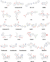
Figure 4. Chemical structures of published small-molecule modulators of CRL activity
Figure 5
Figure 5. Crystal structure of Skp1–Cdc4–SCF-I2 complex (PDB code 3MKS)
Left: the Cdc4–Skp1 protein complex and bound SCF-I2 ligand are shown as molecular surface representations. Right: the expanded inset shows the key residues of Cdc4 (light blue carbons) forming the protein interface that binds SCF-I2 and the ligand chemical structure (yellow carbons); oxygen atoms are in red, and nitrogen atoms are in blue.
Figure 6
Figure 6. Crystal structure of the Skp1–TIR1–Probe 8 complex (PDB code 3C6N)
Left: Skp1–TIR1 protein complex with bound Probe 8 ligand that blocks the interaction of TIR1 with substrate Aux/IAA. Right: the key TIR1 residues (grey carbons) and Probe 8 (yellow carbons) are shown as sticks; oxygen atoms are in red, and nitrogen atoms are in blue.
Figure 7
Figure 7. Crystal structure of the VHL–ElonginB–ElonginC–Compound 7 complex (PDB code 4W9H)
Left: the protein complex and the ligand are shown as molecular surface representations. Right: the insets show the chemical structure of the ligand and a close-up of its binding site, with protein key residues (grey carbons) and ligand molecule (yellow carbons) shown as sticks; oxygen atoms are in red, nitrogen atoms are in blue, and sulfur atoms are in orange.
Figure 8
Figure 8. Crystal structure of the Keap1–SRS-59 complex (PDB code 4L7D)
Left: Keap1 receptor subunit of CRL3Keap1 shown with bound SRS-59 inhibitor that disrupts the interaction with substrate Nrf2. Right: Keap1-binding site with key residues (light blue carbons) and bound SRS-59 molecule (yellow carbons), and the chemical structure of the inhibitor; oxygen atoms are in red; and nitrogen atoms are in blue.
Figure 9
Figure 9. Crystal structure of the Keap1–Cpd16 complex (PDB code 4IQK)
Left: structure Keap1 with bound Cpd16 inhibitor of the Keap1–Nrf2 interaction are shown as molecular surface representations. Right: key residues of Keap1 (light blue carbons) and Cpd16 (yellow carbons) are shown; oxygen atoms are in red, nitrogen atoms are in blue, and sulfur atoms are in orange. Cpd16 binds to the same site as the above mentioned SRS-59.
Figure 10
Figure 10. Crystal structure of the CRBN–DDB1–thalidomide complex (PDB code 4CI1)
Left: CRBN–DDB1 receptor–adaptor complex of CRL4ACRBN with thalidomide bound to CRBN are shown as molecular surface representations. Right: the residues of CRBN (green carbons) forming the interaction interface with thalidomide (yellow carbons); oxygen atoms are in red, and nitrogen atoms are in blue.
Figure 11
Figure 11. Structures of CC0651 bound to an allosteric pocket on the E2-conjugating enzyme Cdc34
Top: crystal structure of the Cdc34–CC0651 binary complex (PDB code 3RZ3). The protein and its ligand are shown as molecular surface representations. Inset: Cdc34 residues (green carbons) forming the binding pocket and the CC0651 ligand (yellow carbons). Bottom: crystal structure of the ternary complex Cdc34A~Ub–CC0651 (PDB code 4MDK). CC0651 (yellow carbons) is bound embedded within the Cdc34A~Ub covalent conjugate protein. CC0651 suppresses the hydrolysis of the thioester bond between the catalytic cysteine residue of Cdc34 (green carbons, cysteine residue not shown) and ubiquitin (brown carbons, Lys48 side chain shown). Oxygen atoms are in red, nitrogen atoms are in blue, and chlorine atoms are in light green.
Figure 12
Figure 12. Crystal structure of the NAE1–UBA3–NEDD8~MLN4924 complex (PDB code 3GZN)
The NAE1 regulatory subunit and the UBA3 catalytic subunit form the heterodimeric NAE. Left: NAE complexed with NEDD8~MLN4924 covalent adduct, where MLN4924 inhibits the active site of NAE. Right: the residues forming interface between NAE1 (grey carbons), UBA3 (green carbons), NEDD8 (cyan carbons) and MLN4924 (yellow carbons). Oxygen atoms are in red, nitrogen atoms are in blue, and sulfur atoms are in orange.

References

    1. Ciechanover A., Hershko A., Rose I. 2004. The Nobel Prize in Chemistry 2004, http://www.nobelprize.org/nobel_prizes/chemistry/laureates/2004/ - PMC - PubMed
    1. Pickart C. M. Mechanisms underlying ubiquitination. Annu. Rev. Biochem. 2001;70:503–533. doi: 10.1146/annurev.biochem.70.1.503. - DOI - PubMed
    1. Hershko A., Ciechanover A. The ubiquitin system. Annu. Rev. Biochem. 1998;67:425–479. doi: 10.1146/annurev.biochem.67.1.425. - DOI - PubMed
    1. Komander D. The emerging complexity of protein ubiquitination. Biochem. Soc. Trans. 2009;37:937–953. doi: 10.1042/BST0370937. - DOI - PubMed
    1. Rubin D. M., Finley D. Proteolysis. The proteasome: a protein-degrading organelle? Curr. Biol. 1995;5:854–858. doi: 10.1016/S0960-9822(95)00172-2. - DOI - PubMed

Publication types