. 2015 Apr 17;10(4):e0124487.
doi: 10.1371/journal.pone.0124487. eCollection 2015.

The 15kDa selenoprotein and thioredoxin reductase 1 promote colon cancer by different pathways

Affiliations

The 15kDa selenoprotein and thioredoxin reductase 1 promote colon cancer by different pathways

Petra A Tsuji et al. PLoS One. .

Abstract

Selenoproteins mediate much of the cancer-preventive properties of the essential nutrient selenium, but some of these proteins have been shown to also have cancer-promoting effects. We examined the contributions of the 15kDa selenoprotein (Sep15) and thioredoxin reductase 1 (TR1) to cancer development. Targeted down-regulation of either gene inhibited anchorage-dependent and anchorage-independent growth and formation of experimental metastases of mouse colon carcinoma CT26 cells. Surprisingly, combined deficiency of Sep15 and TR1 reversed the anti-cancer effects observed with down-regulation of each single gene. We found that inflammation-related genes regulated by Stat-1, especially interferon-γ-regulated guanylate-binding proteins, were highly elevated in Sep15-deficient, but not in TR1-deficient cells. Interestingly, components of the Wnt/β-catenin signaling pathway were up-regulated in cells lacking both TR1 and Sep15. These results suggest that Sep15 and TR1 participate in interfering regulatory pathways in colon cancer cells. Considering the variable expression levels of Sep15 and TR1 found within the human population, our results provide insights into new roles of selenoproteins in cancer.


Conflict of interest statement

Competing Interests: The authors have declared that no competing interests exist.

Figures

Fig 1
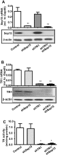
Fig 1. Expression of Sep15 and TR1 in CT26 cells.
Cells were stably transfected with the pU6-m3 control, shSep15, shTR1 or shTR1/shSep15 (as indicated). (A) Expression of Sep15 by real-time RT-PCR (upper panel) and Western blotting (lower panel). (B) Expression of TR1 by real-time RT-PCR (upper panel) and Western blotting (lower panel). (C) Thioredoxin reductase activity. Columns, mean (n = 3–6); bars, SE; (*P<0.05, **P<0.01, compared to control).
Fig 2
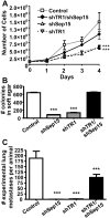
Fig 2. Anchorage-dependent, -independent growth and formation of lung metastases.
(A) Growth rates of shSep15, shTR1 and shTR1/Sep15 cells compared to controls (n = 6). (B) Anchorage-independent growth in soft agar of shSep15, shTR1 and shTR1/Sep15 cells compared to controls (n = 4–8). (C) Formation of experimental lung metastases after i.v. injection of 5×10⁵ cells (control, shSep15, shTR1, or shTR1/shSep15) into BALB/c mice (n = 10/construct). (***P<0.001, compared to control).
Fig 3
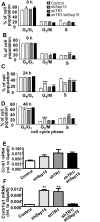
Fig 3. Cell cycle analysis.
Percent of cells in each cell cycle phase as determined by FACS analysis at (A) 0 h; (B) 6 h; (C) 24 h; and (D) 48 h after release from cell synchronization. mRNA expression of (E) CyclinB1 (Ccnb1), and (F) CyclinB1 Interacting-Protein-1 (Ccnb1ip1), as determined by real-time RT-PCR. Columns, mean (n = 3–9); bars, SE; (*P<0.05, **P<0.01, ***P<0.001).
Fig 4
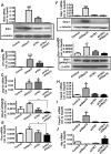
Fig 4. Validation of gene expression for genes significantly changed in shSep15 cells.
Expression of (A) Ifi44 mRNA (upper panel) and protein (lower panel); (B) Irf-7 mRNA; (C) Usp18 mRNA; (D) Gbp-6 mRNA; (E) Ifnγ mRNA; (F) Gbp-1 mRNA (upper panel) and protein (lower panel); (G) Stat-1 mRNA (upper panel) and protein (lower panel); (H), Casp6 mRNA; (I) Casp12 mRNA; and (J) Afp mRNA. Expression of mRNA in control, shSep15, shTR1, and shTR1/Sep15 cells was determined by real-time RT-PCR, and expressed relative to Gapdh. Columns, mean (n = 3–6); bars, SE; (*P<0.05, **P<0.01, ***P<0.001). Protein expression was determined by Western blotting, and expressed relative to β-actin or α-tubulin, as indicated.
Fig 5
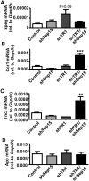
Fig 5. Possible connection to Wnt/β-catenin signaling pathway in shTR1/Sep15 cells.
mRNA levels of (A) Speg; (B) Ccr1; and (C) Tnc; and (D) Apc in control, shSep15, shTR1, and shTR1/Sep15 cells, as determined by real-time RT-PCR, and expressed relative to Gapdh. Columns, mean (n = 3–6); bars, SE; (**P<0.01, ***P<0.001).
Fig 6
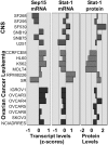
Fig 6. Signatures of selected NCI-60 cell lines for Sep15 and Stat-1.
Opposing directions of transcript intensity scores (z-scores) for Sep15 and Stat-1 mRNAs were highly significant (r = -0.646, P<0.01) for human central nervous system (CNS), leukemia and ovarian cancer cell lines. Mean centered average protein activity patterns for Stat-1 (Stat-1_20 antibody) significantly correlated (r = 0.661, P<0.01) with Stat-1 mRNA expression.

References

    1. American Cancer Society. Cancer Facts & Figures 2013. Atlanta: American Cancer Society, 2013.
    1. Duffield-Lillico AJ, Dalkin BL, Reid ME, Turnbull BW, Slate EH, Jacobs ET, et al. Selenium supplementation, baseline plasma selenium status and incidence of prostate cancer: an analysis of the complete treatment period of the Nutritional Prevention of Cancer Trial. BJU Int. 2003;91: 608–612. - PubMed
    1. Jao SW, Shen KL, Lee WP, Ho YS. Effect of selenium on 1,2-dimethylhydrazine-induced intestinal cancer in rats. Dis Colon Rectum. 1996;39: 628–631. - PubMed
    1. Almondes KG, Leal GV, Cozzolino SM, Philippi ST, Rondó PH. The role of selenoproteins in cancer. Rev Assoc Med Bras. 2010;56: 484–488. - PubMed
    1. Apostolou S, Klein JO, Mitsuuchi Y, Shetler JN, Poulikakos PL, Jhanwar SC, et al. Growth inhibition and induction of apoptosis in mesothelioma cells by selenium and dependence on selenoprotein SEP15 genotype. Oncogene. 2004;23: 5032–5040. - PubMed

Publication types

Associated data