. 2015 Apr 10:12:69.
doi: 10.1186/s12974-015-0289-5.

Attenuation of traumatic brain injury-induced cognitive impairment in mice by targeting increased cytokine levels with a small molecule experimental therapeutic

Affiliations

Attenuation of traumatic brain injury-induced cognitive impairment in mice by targeting increased cytokine levels with a small molecule experimental therapeutic

Adam D Bachstetter et al. J Neuroinflammation. .

Abstract

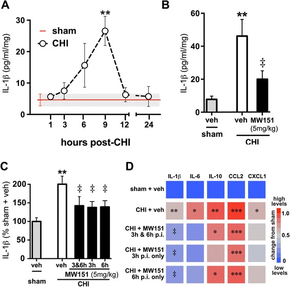
Background: Evidence from clinical studies and preclinical animal models suggests that proinflammatory cytokine overproduction is a potential driving force for pathology progression in traumatic brain injury (TBI). This raises the possibility that selective targeting of the overactive cytokine response, a component of the neuroinflammation that contributes to neuronal dysfunction, may be a useful therapeutic approach. MW151 is a CNS-penetrant, small molecule experimental therapeutic that selectively restores injury- or disease-induced overproduction of proinflammatory cytokines towards homeostasis. We previously reported that MW151 administered post-injury (p.i.) is efficacious in a closed head injury (CHI) model of diffuse TBI in mice. Here we test dose dependence of MW151 to suppress the target mechanism (proinflammatory cytokine up-regulation), and explore the therapeutic window for MW151 efficacy.

Methods: We examined suppression of the acute cytokine surge when MW151 was administered at different times post-injury and the dose-dependence of cytokine suppression. We also tested a more prolonged treatment with MW151 over the first 7 days post-injury and measured the effects on cognitive impairment and glial activation.

Results: MW151 administered up to 6 h post-injury suppressed the acute cytokine surge, in a dose-dependent manner. Administration of MW151 over the first 7 days post-injury rescues the CHI-induced cognitive impairment and reduces glial activation in the focus area of the CHI.

Conclusions: Our results identify a clinically relevant time window post-CHI during which MW151 effectively restores cytokine production back towards normal, with a resultant attenuation of downstream cognitive impairment.


Figures

Figure 1
Figure 1
Time window for suppression of acute cytokine surge by MW151. (A) Temporal changes in IL-1β protein levels were measured in tissue homogenates from the cortex of adult C57BL/6 J mice subjected to sham conditions (red line mean ± SEM), or at select time points after the CHI (n = 4 per group). (B) Treatment of mice with MW151 (5 mg/kg i.p.) at 3 and 6 h post-injury (p.i.) significantly reduced IL-1β up-regulation at 9 h p.i., compared to CHI + saline vehicle (veh) control (n = 4 sham; n = 9 CHI + veh; n = 10 CHI + MW151). (C, D) Injured mice were treated with veh or MW151 (5 mg/kg i.p.) at 3 h and 6 h, at 3 h only, or at 6 h only, and cytokine and chemokine protein levels were measured in cortex homogenates at 9 h p.i. (n = 7 to 20 per group). (C) Injury-induced IL-1β levels were significantly suppressed with MW151 regardless of time of treatment. (D) Heatmap of the relative changes in cytokine and chemokine protein levels showing the specificity of MW151 for IL-1β and IL-6. Significance for a given inflammatory marker is indicated by *P < 0.05, **P < 0.005, ***P < 0.001 compared to sham treated mice. ‡P < 0.05 compared to CHI + veh.
Figure 2
Figure 2
Dose-dependent suppression of IL-1β up-regulation by MW151. Mice were subjected to sham injury or CHI, and injected i.p. at 6 h p.i. with saline vehicle (veh). CHI mice were injected i.p. at 6 h p.i. with MW151 at one of three different doses (1, 5, and 10 mg/kg). Levels of IL-1β (A) and CCL2 (B) protein were measured in cortex homogenates at the 9-h p.i. time point. *P < 0.05, ***P < 0.001 compared to sham mice. ‡P < 0.05, ‡‡P < 0.01 CHI + veh compared to CHI + MW151 (n = 10 sham + veh; n = 20 CHI + veh; n = 9 to 10 CHI + MW151).
Figure 3
Figure 3
Functional efficacy of MW151 to suppress CHI-induced cognitive impairment. (A) Overview of study design. MW151 (5 mg/kg i.p.) was administered either as a single acute treatment at 6 h p.i. or in a more extended treatment paradigm (3 h, 6 h, then once daily for 7 days p.i.). (B) Mice were tested for cognitive deficits in the RAWM starting at 14 days p.i. (n = 23 sham; n = 27 CHI + veh; n = 12 CHI + MW150 acute; n = 14 CHI + MW150 chronic).
Figure 4
Figure 4
Effects of MW151 treatment on glia morphological activation. Mice were subjected to sham injury or CHI and administered veh control; injured mice were administered MW151 (5 mg/kg i.p.) at 3 h, 6 h, and once daily for 7 days p.i.. Brain tissue was prepared for IHC at 16 days p.i. and stained for microglia (IBA1, CD45) and astrocyte (GFAP) morphological activation. (A) Overview of regions of interest (ROIs) used for quantitative neuropathological analysis of glia activation using the Aperio ScanScope. Heatmap shows the effect of injury and MW151 treatment in five ROIs for IBA1 (B), CD45 (C), and GFAP (D) IHC. Treatment with MW151 reduced IBA1, CD45, and GFAP staining in the focus region of the cortex. There was little effect of MW151 treatment on glial staining in other areas of the brain. **P < 0.005 compared to sham mice. See also Table 2. Representative photomicrographs of IBA1 (E), CD45 (F), and GFAP (G) in the focus region of the neocortex show the robust glial morphological activation that occurs in response to the injury.

References

    1. Faul M, Xu L, Wald MM, Coronado VG. Traumatic brain injury in the United States: emergency department visits, hospitalizations, and deaths. Centers for Disease Control and Prevention, National Center for Injury Prevention and Control Atlanta (GA): 2010.
    1. Bazarian JJ, Cernak I, Noble-Haeusslein L, Potolicchio S, Temkin N. Long-term neurologic outcomes after traumatic brain injury. J Head Trauma Rehabil. 2009;24(6):439–51. doi: 10.1097/HTR.0b013e3181c15600. - DOI - PubMed
    1. Jordan BD. Chronic traumatic encephalopathy and other long-term sequelae. Continuum (Minneap Minn) 2014;20(6 Sports Neurology):1588–604. - PubMed
    1. Ramlackhansingh AF, Brooks DJ, Greenwood RJ, Bose SK, Turkheimer FE, Kinnunen KM, et al. Inflammation after trauma: microglial activation and traumatic brain injury. Ann Neurol. 2011;70(3):374–83. doi: 10.1002/ana.22455. - DOI - PubMed
    1. Heneka MT, Kummer MP, Latz E. Innate immune activation in neurodegenerative disease. Nat Rev Immunol. 2014;14(7):463–77. doi: 10.1038/nri3705. - DOI - PubMed

Publication types

MeSH terms