. 2015 Mar 24:12:27.
doi: 10.1186/s12977-015-0156-6.

Modulation of human endogenous retrovirus (HERV) transcription during persistent and de novo HIV-1 infection

Affiliations

Modulation of human endogenous retrovirus (HERV) transcription during persistent and de novo HIV-1 infection

Michelle Vincendeau et al. Retrovirology. .

Abstract

Background: The human genome contains multiple LTR elements including human endogenous retroviruses (HERVs) that together account for approximately 8-9% of the genomic DNA. At least 40 different HERV groups have been assigned to three major HERV classes on the basis of their homologies to exogenous retroviruses. Although most HERVs are silenced by a variety of genetic and epigenetic mechanisms, they may be reactivated by environmental stimuli such as exogenous viruses and thus may contribute to pathogenic conditions. The objective of this study was to perform an in-depth analysis of the influence of HIV-1 infection on HERV activity in different cell types.

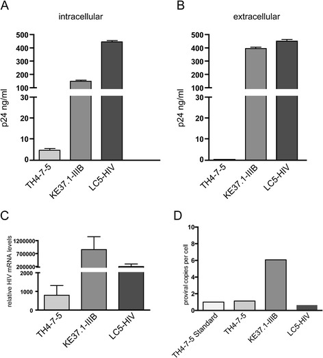
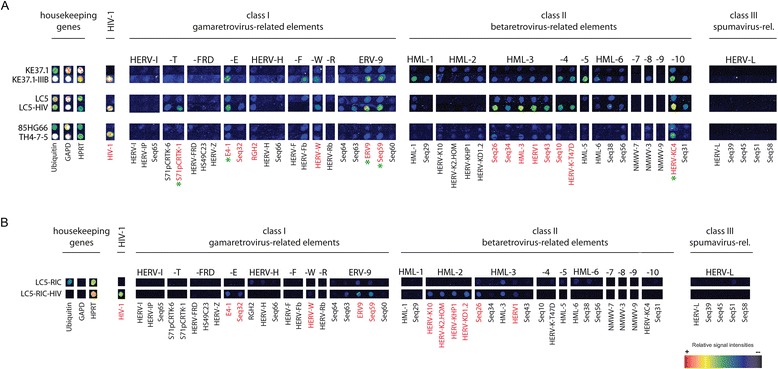
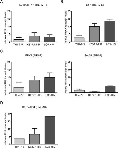
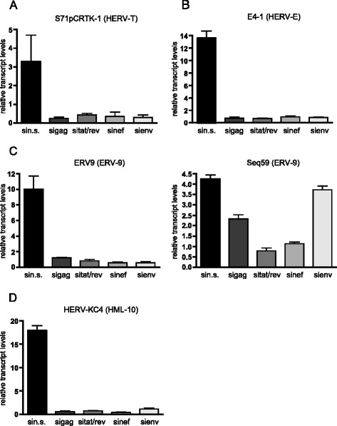
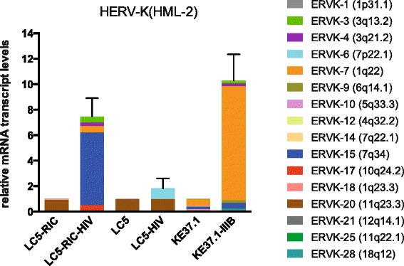
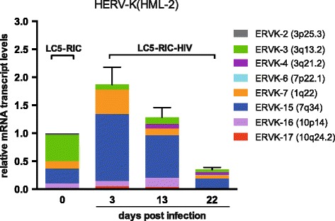
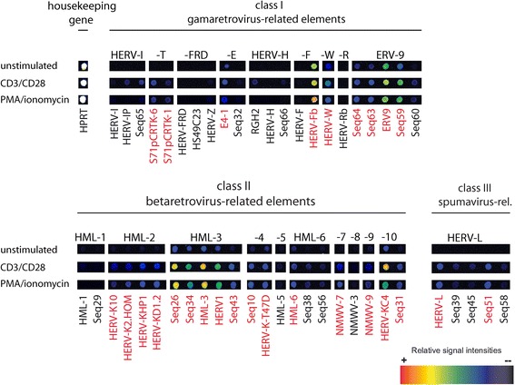
Results: A retrovirus-specific microarray that covers major HERV groups from all three classes was used to analyze HERV transcription patterns in three persistently HIV-1 infected cell lines of different cellular origins and in their uninfected counterparts. All three persistently infected cell lines showed increased transcription of multiple class I and II HERV groups. Up-regulated transcription of five HERV taxa (HERV-E, HERV-T, HERV-K (HML-10) and two ERV9 subgroups) was confirmed by quantitative reverse transcriptase PCR analysis and could be reversed by knock-down of HIV-1 expression with HIV-1-specific siRNAs. Cells infected de novo by HIV-1 showed stronger transcriptional up-regulation of the HERV-K (HML-2) group than persistently infected cells of the same origin. Analysis of transcripts from individual members of this group revealed up-regulation of predominantly two proviral loci (ERVK-7 and ERVK-15) on chromosomes 1q22 and 7q34 in persistently infected KE37.1 cells, as well as in de novo HIV-1 infected LC5 cells, while only one single HML-2 locus (ERV-K6) on chromosome 7p22.1 was activated in persistently infected LC5 cells.

Conclusions: Our results demonstrate that HIV-1 can alter HERV transcription patterns of infected cells and indicate a correlation between activation of HERV elements and the level of HIV-1 production. Moreover, our results suggest that the effects of HIV-1 on HERV activity may be far more extensive and complex than anticipated from initial studies with clinical material.


Figures

Figure 1
Figure 1
Productivity of different persistently HIV-1 infected human cell lines. (A) Intracellular p24-production in the infected cell lines TH4-7-5, KE37.1-IIIB and LC5-HIV. (B) Extracellular p24-production in the infected cell lines TH4-7-5, KE37.1-IIIB and LC5-HIV. Quantification of p24 antigen in infected cells and in the supernatant was performed as described [82] using a p24 ELISA. (C) HIV-1 transcript levels in the persistently infected cell lines TH4-7-5, KE37.1-IIIB and LC5-HIV. (D) Provirus copy numbers of the infected cell lines TH4-7-5, KE37.1-IIIB and LC5-HIV measured with quantitative PCR. Absolute quantification of HIV proviral copies was performed as described in [82].
Figure 2
Figure 2
HERV transcription profiles of HIV-1 infected cell lines examined by a retrovirus-specific microarray. False color mapping was used for image visualization. The house keeping genes ubiquitin, glycerinaldehyde-3-phosphate-dehydrogenase (GAPDH) and hypoxanthine-guanine phosphoribosyltransferase (HPRT) served as a quality control and internal standard. HIV-1 oligonucleotides are also spotted on the chip as a positive control and to demonstrate HIV-1 infection. HERVs are grouped in class I, II and III elements. It should be noted that each positive spot on the microarray can represent multiple HERV proviruses of one multicopy HERV subgroup with sufficient sequence similarity that individual elements cannot be distinguished. (A) Comparison of persistently HIV-1 infected cell lines with the corresponding uninfected cell lines. Up-regulated HERV subgroups are indicated by red letters. HERV subgroups marked with a green asterisk were additionally analyzed by qRT-PCR. (B) Comparison of de novo HIV-1 infected LC5-RIC cells with uninfected cells.
Figure 3
Figure 3
Relative transcriptional activity of selected HERV subgroups. Relative transcript levels of (A) HERV taxa S71pCRTK-1 (group HERV-T), (B) E4-1 (group HERV-E), (C) ERV9 and Seq59 (both group ERV9) and (D) HERV-KC4 (group HERV-K (HML-10)) were determined by qRT-PCR. The Y-axis shows the x-fold relative expression of HERV-transcripts in infected cells referred to uninfected cells. Relative transcription was quantified according to [85] and normalized to RNA Polymerase II (RPII) transcript levels. Standard errors for triplicate experiments are indicated.
Figure 4
Figure 4
Down-regulation of HIV-1 induced HERV activity by siRNAs targeting HIV-1 transcripts. LC5-HIV cells were treated with the RNAiFect transfection reagent (mock), non-silencing siRNAs (sin.s.) or with siRNAs against gag (sigag), rev (sirev), nef (sinef) and env (sienv). Relative transcript levels of (A) HERV taxa S71pCRTK-1 (group HERV-T), (B) E4-1 (group HERV-E), (C) ERV9 and Seq59 (both group ERV9) and (D) HERV-KC4 (group HERV-K (HML-10)) were determined by qRT-PCR. The Y-axis shows the x-fold relative HERV transcript levels in LC5-HIV cells transfected with non-silencing siRNA (sin.s) and HIV-1-specific siRNAs (sigag, sitat/rev, sinef, sienv) referred to uninfected control cells. The data were normalized to the house-keeping gene RNA Polymerase II (RPII). Standard errors for triplicate experiments are indicated.
Figure 5
Figure 5
Transcriptional activity of HERV-K (HML-2) proviral loci in de novo and persistently infected cells. The Y-axis shows the x-fold relative expression of HERV-K (HML-2) gag transcripts in infected cells referred to uninfected cells. Relative transcription was quantified according to [85] and normalized to RPII transcript levels. HERV-K (HML-2) transcripts were amplified using gag-specific primers, cloned, sequenced and assigned to proviral loci as described previously [58]. For each HERV-K (HML-2) locus the relative cloning frequency of cDNA is shown as percentage of the total HERV-K (HML-2) transcription level determined by qRT-PCR.
Figure 6
Figure 6
Time course of HERV-K (HML-2) in de novo HIV-1 infected LC5-RIC cells. The Y-axis shows the x-fold relative expression of HERV-K (HML-2) gag transcripts in infected cells referred to uninfected cells. RNA was isolated from LC5-RIC cells prior to infection and from cells infected with HIV-1IIIB/LAI at day 3, 13 and 22. Relative transcription was quantified according to [85] and normalized to RPII transcript levels. HERV-K (HML-2) transcripts were amplified using gag-specific primers, cloned, sequenced and assigned to proviral loci as described previously [58]. For each HERV-K (HML-2) locus the relative cloning frequency of cDNA is shown as percentage of the total HERV-K (HML-2) transcription level determined by qRT-PCR.
Figure 7
Figure 7
HERV transcription profiles of PMA/ionomycin and CD3/CD28 stimulated Jurkat T-cells. False color mapping was used for image visualization. The house keeping gene HPRT served as a quality control and internal standard. Up-regulated HERV subgroups are indicated by red letters.

References

    1. Sverdlov E. Retroviruses and primate genome evolution. Georgetown, Texas, USA: Landes Bioscience; 2005.
    1. Belshaw R, Katzourakis A, Paces J, Burt A, Tristem M. High copy number in human endogenous retrovirus families is associated with copying mechanisms in addition to reinfection. Mol Biol Evol. 2005;22:814–7. doi: 10.1093/molbev/msi088. - DOI - PubMed
    1. Belshaw R, Pereira V, Katzourakis A, Talbot G, Paces J, Burt A, et al. Long-term reinfection of the human genome by endogenous retroviruses. Proc Natl Acad Sci U S A. 2004;101:4894–9. doi: 10.1073/pnas.0307800101. - DOI - PMC - PubMed
    1. Mager DL, Medstrand P. Retroviral repeat sequences. In: Cooper D, editor. Nature encyclopedia of the human genome. D. Cooper edition. London, United Kingdom: Nature Publishing Group; 2003. pp. 57–63.
    1. Evans LH, Alamgir AS, Owens N, Weber N, Virtaneva K, Barbian K, et al. Mobilization of endogenous retroviruses in mice after infection with an exogenous retrovirus. J Virol. 2009;83:2429–35. doi: 10.1128/JVI.01926-08. - DOI - PMC - PubMed