

Peritumoral edema shown by MRI predicts poor clinical outcome in glioblastoma
- PMID: 25886608
- PMCID: PMC4358863
- DOI: 10.1186/s12957-015-0496-7
Peritumoral edema shown by MRI predicts poor clinical outcome in glioblastoma
Abstract
Background: Magnetic resonance imaging (MRI) plays an irreplaceable role in the preoperative diagnosis of glioma, and its imaging features are the base of making treatment decisions in patients with glioma, but it is still controversial whether peritumoral edema shown by MRI from preoperative routine scans are associated with patient survival. The aim of this study was to assess the prognostic value of preoperative MRI features in patients with glioblastoma.
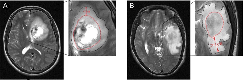
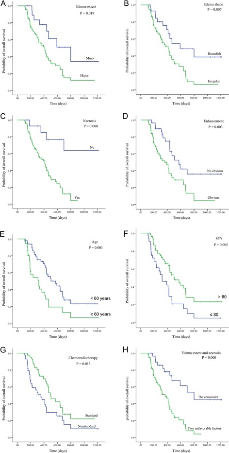
Methods: A retrospective review of 87 patients with newly diagnosed supratentorial glioblastoma was performed using medical records and MRI data from routine scans. The Kaplan-Meier method and COX proportional hazard model were applied to evaluate the prognostic impact on overall survival of pretreatment MRI features (including peritumoral edema, edema shape, necrosis, cyst, enhancement, tumor crosses midline, edema crosses midline, and tumor size).
Results: In addition to patient age, Karnofsky performance status (KPS) and postoperative chemoradiotherapy, peritumoral edema extent and necrosis on preoperative MRI were independent prognostic indicator for poor survival. Furthermore, patients with two unfavorable conditions (major edema and necrosis) had a shorter overall survival compared with the remainder.
Conclusions: Our data confirm that peritumoral edema extent and necrosis are helpful for predicting poor clinical outcome in glioblastoma. These features were easy to determine from routine MRI scans postoperatively and therefore could provide a certain instructive significance for clinical activities.
Figures
References
-
- Central Brain Tumor Registry of the United States . CBTRUS statistical report: primary brain and central nervous system tumors in the United States in 2004–2006. Hinsdale, Ill: Central Brain Tumor Registry of the United States; 2010.
-
- Dahlrot RH. The prognostic value of clinical factors and cancer stem cell-related markers in gliomas. Dan Med J. 2014;61(10):B4944. - PubMed
Publication types
MeSH terms
LinkOut - more resources
Full Text Sources
Other Literature Sources
Medical
