. 2015 Apr 11:10:16.
doi: 10.1186/s13062-015-0050-0.

Sequence analysis reveals a conserved extension in the capping enzyme of the alphavirus supergroup, and a homologous domain in nodaviruses

Affiliations

Sequence analysis reveals a conserved extension in the capping enzyme of the alphavirus supergroup, and a homologous domain in nodaviruses

Tero Ahola et al. Biol Direct. .

Abstract

Background: Members of the alphavirus supergroup include human pathogens such as chikungunya virus, hepatitis E virus and rubella virus. They encode a capping enzyme with methyltransferase-guanylyltransferase (MTase-GTase) activity, which is an attractive drug target owing to its unique mechanism. However, its experimental study has proven very difficult.

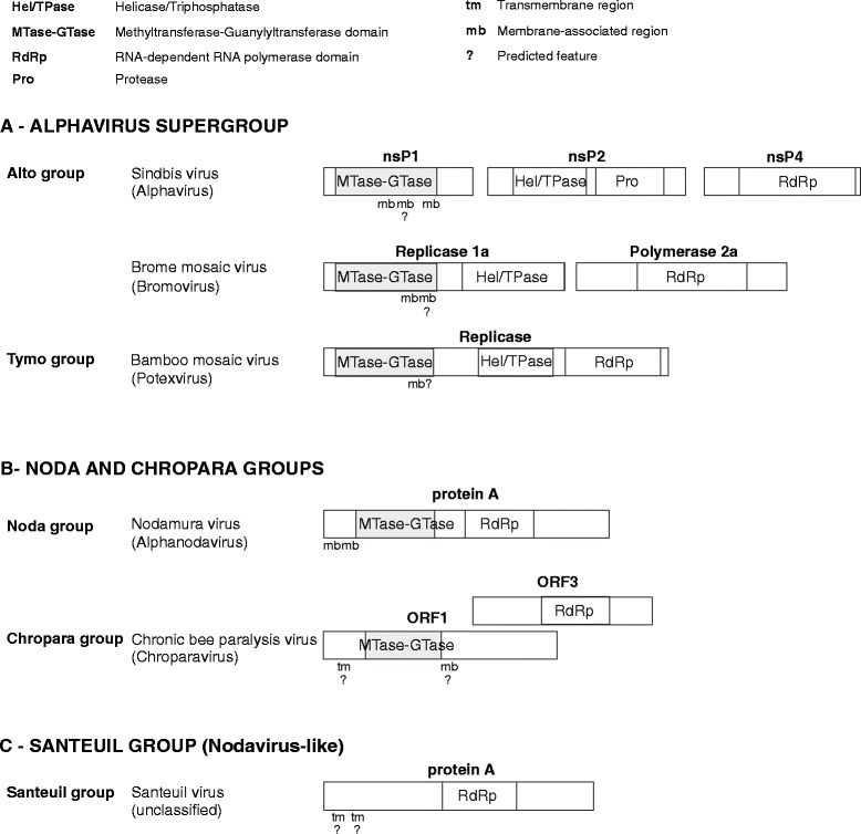
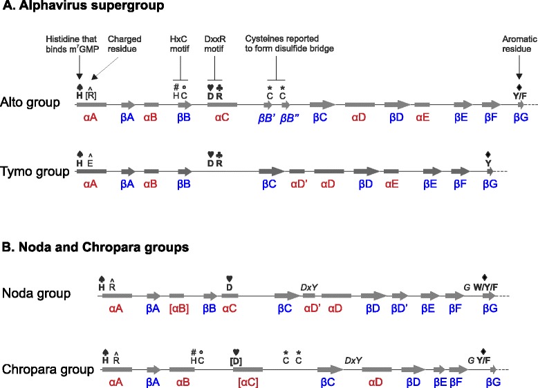
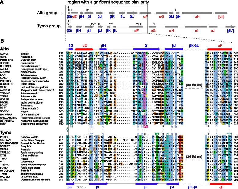
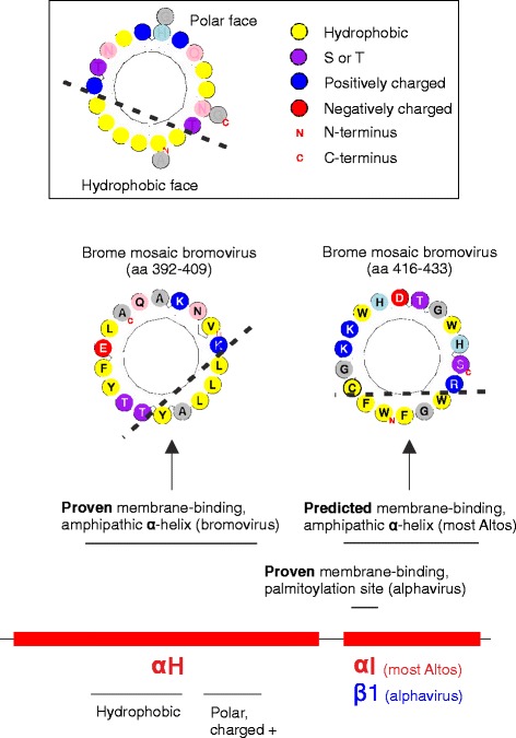
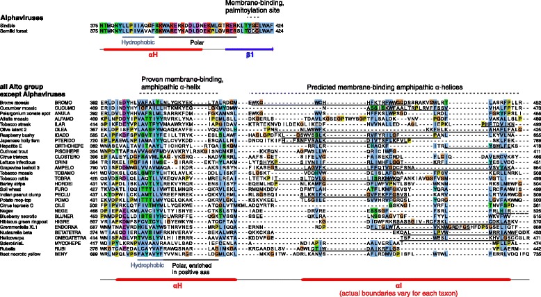
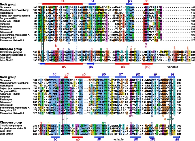
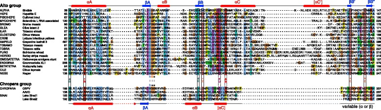
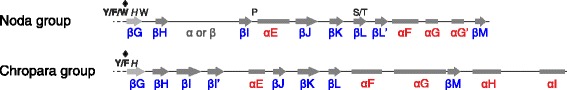
Results: We examined over 50 genera of viruses by sequence analyses. Earlier studies showed that the MTase-GTase contains a "Core" region conserved in sequence. We show that it is followed by a long extension, which we termed "Iceberg" region, whose secondary structure, but not sequence, is strikingly conserved throughout the alphavirus supergroup. Sequence analyses strongly suggest that the minimal capping domain corresponds to the Core and Iceberg regions combined, which is supported by earlier experimental data. The Iceberg region contains all known membrane association sites that contribute to the assembly of viral replication factories. We predict that it may also contain an overlooked, widely conserved membrane-binding amphipathic helix. Unexpectedly, we detected a sequence homolog of the alphavirus MTase-GTase in taxa related to nodaviruses and to chronic bee paralysis virus. The presence of a capping enzyme in nodaviruses is biologically consistent, since they have capped genomes but replicate in the cytoplasm, where no cellular capping enzyme is present. The putative MTase-GTase domain of nodaviruses also contains membrane-binding sites that may drive the assembly of viral replication factories, revealing an unsuspected parallel with the alphavirus supergroup.

Conclusions: Our work will guide the functional analysis of the alphaviral MTase-GTase and the production of domains for structure determination. The identification of a homologous domain in a simple model system, nodaviruses, which replicate in numerous eukaryotic cell systems (yeast, flies, worms, mammals, and plants), can further help crack the function and structure of the enzyme.


Figures

Figure 1
Figure 1
Organization of the replicase domains in the viral groups studied. The representation is approximately to scale, and incorporates the findings made in this study. See graphical legend (top) for abbreviations. Many members of the alphavirus supergroup encode a polyprotein, which is cleaved into several proteins or expressed as individual proteins, as exemplified by Sindbis virus and brome mosaic virus, respectively. In contrast, the BaMV replicase is not cleaved. For each virus, only proteins or domains containing a MTase-GTase, a protease, an RdRp, or a triphosphatase are shown. Membrane association segments (mb) that are predicted herein, rather than experimentally determined, are indicated by “?”.
Figure 2
Figure 2
Organization of the Core region of the MTase-GTase. The representation is approximately to scale. α-helices and β-strands are indicated by rectangles and arrows, respectively. The charged residue (R^ or E^) in helix αA is located 7aas downstream of the histidine H♠, and the two cysteines (C*) upstream of βC are spaced by 6 aas. Secondary structure elements or positions that are not strictly conserved are indicated by brackets. Numbering of secondary elements in the noda and chropara groups is as for the alphavirus supergroup. The conserved positions unique to the noda and chropara groups are in italics.
Figure 3
Figure 3
The Iceberg region, downstream the MTase-GTase Core in the alphavirus supergroup. A. Consensus secondary structure of the Iceberg region (which starts after the conserved Y♦ in strand βG) for the alto and tymo groups. Conventions are the same as in Figure 2. Residues conserved in each group are indicated. B. Sequence alignment of the region boxed in A, with the ClustalX coloring scheme [96]. The sequences of the tymo group were aligned to the set alignment of the alto group by using MAFFT with the --add option [94]. Substituted residues Y299 and W222 of BaMV are in bold (see text). Abbreviations: p, polar; h, hydrophobic; s, small.
Figure 4
Figure 4
Overview of the membrane-binding regions in the MTase-GTase of the alto group. Known or predicted membrane association sites are indicated above the C-terminus of the Iceberg region of the alto group, with the same conventions as in Figure 3A. Amphipathic helices are depicted in helical wheel representation.
Figure 5
Figure 5
Sequences of the C-terminus of the Iceberg region of the alto group, with known and predicted membrane-binding regions. Sequence alignment of the last two secondary structure elements of the Iceberg region of the alto group. Conventions are the same as in Figure 3. The penultimate secondary element is αH in all taxa, and the last element is either a β-strand (β1), in alphaviruses, or an α-helix (αI) in other genera (see also Figure 4). The experimentally characterized, amphipathic helix of bromoviruses is doubly underlined. Amphipathic helices predicted by Heliquest are singly underlined. The sequences of each genus have no significant sequence similarity, and were aligned instead according to their secondary structure and hydrophobicity, using Promals [39].
Figure 6
Figure 6
Comparison between the MTase-GTase Core of the noda and chropara groups. Conventions are the same as in Figures 2 and 3. Numbering of secondary elements is as for the alphavirus supergroup, and by analogy, the Core region ends at the position W/Y/F♦ in strand βG. The sequences of the chropara group were aligned to the set alignment of the noda group by using MAFFT with the --add option [94]. Conserved cysteines in the chropara group, which may be equivalent to those of the alto group, are indicated by an asterisk. The putative equivalent of the conserved D♥ of the alphavirus supergroup is boxed with a dashed line.
Figure 7
Figure 7
Parallel between the MTase-GTase of the alto and chropara groups. Conventions are the same as in Figures 2, 3 and 6. The sequences of the chropara group were aligned to the set alignment of the alto group by using MAFFT with the --add option [94].
Figure 8
Figure 8
C-terminal extension (Iceberg) of the Core MTase-GTase in the noda and chropara groups. Conventions are the same as in Figures 2 and 3. The C-terminal extension starts after the conserved position W/Y/F♦ in strand βG.

References

    1. Goldbach R. Genome similarities between plant and animal RNA viruses. Microbiol Sci. 1987;4(7):197–202. - PubMed
    1. Koonin EV, Dolja VV. Evolution and taxonomy of positive-strand RNA viruses: implications of comparative analysis of amino acid sequences. Crit Rev Biochem Mol Biol. 1993;28(5):375–430. doi: 10.3109/10409239309078440. - DOI - PubMed
    1. Salonen A, Ahola T, Kaariainen L. Viral RNA replication in association with cellular membranes. Curr Top Microbiol Immunol. 2005;285:139–73. - PMC - PubMed
    1. Firth AE, Brierley I. Non-canonical translation in RNA viruses. J Gen Virol. 2012;93(Pt 7):1385–409. doi: 10.1099/vir.0.042499-0. - DOI - PMC - PubMed
    1. Decroly E, Ferron F, Lescar J, Canard B. Conventional and unconventional mechanisms for capping viral mRNA. Nat Rev Microbiol. 2012;10(1):51–65. - PMC - PubMed

Publication types

MeSH terms

LinkOut - more resources