Clinical Trial
. 2015 Apr 16:15:33.
doi: 10.1186/s12894-015-0026-5.

Prostatic arterial embolization for the treatment of lower urinary tract symptoms due to large (>80 mL) benign prostatic hyperplasia: results of midterm follow-up from Chinese population

Affiliations
Clinical Trial

Prostatic arterial embolization for the treatment of lower urinary tract symptoms due to large (>80 mL) benign prostatic hyperplasia: results of midterm follow-up from Chinese population

Mao Qiang Wang et al. BMC Urol. .

Abstract

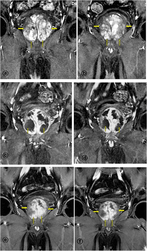
Background: Currently, large prostate size (>80 mL) of benign prostatic hyperplasia (BPH) still pose technical challenges for surgical treatment. This prospective study was designed to explore the safety and efficacy of prostatic arterial embolization (PAE) as an alternative treatment for patients with lower urinary tract symptoms (LUTS) due to largeBPH.

Methods: A total of 117 patients with prostates >80 mL were included in the study; all were failure of medical treatment and unsuited for surgery. PAE was performed using combination of 50-μm and 100-μm particles in size, under local anaesthesia by a unilateral femoral approach. Clinical follow-up was performed using the international prostate symptoms score (IPSS), quality of life (QoL), peak urinary flow (Qmax), post-void residual volume (PVR), international index of erectile function short form (IIEF-5), prostatic specific antigen (PSA) and prostatic volume (PV) measured by magnetic resonance (MR) imaging, at 1, 3, 6 and every 6 months thereafter.

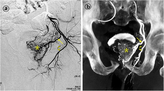
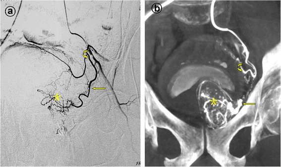
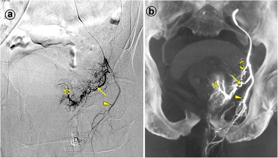
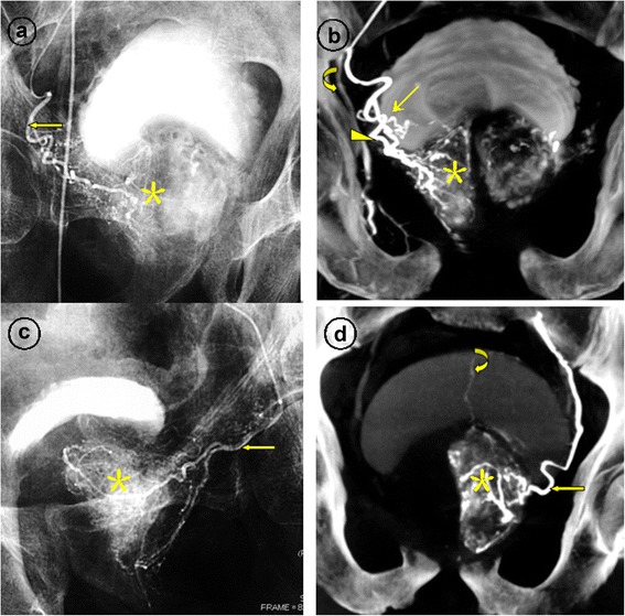
Results: The prostatic artery origins in this study population were different from previously published results. PAE was technically successful in 109 of 117 patients (93.2%). Follow-up data were available for the 105 patients with a mean follow-up of 24 months. The clinical improvements in IPSS, QoL, Qmax, PVR, and PV at 1, 3, 6, 12, and 24 months was 94.3%, 94.3%, 93.3%, 92.6%, and 91.7%, respectively. The mean IPSS (pre-PAE vs post-PAE 26.0 vs 9.0; P < .0.01), the mean QoL (5.0 vs 3.0; P < 0.01), the mean Qmax (8.5 vs 14.5; P < 0.01), the mean PVR (125.0 vs 40.0; P < 0.01), and PV (118.0 vs 69.0, with a mean reduction of 41.5%; P < 0.01 ) at 24-month after PAE were significantly different with respect to baseline. The mean IIEF-5 was not statistically different from baseline. No major complications were noted.

Conclusions: PAE is a safe and effective treatment method for patients with LUTS due to large volume BPH. PAE may play an important role in patients in whom medical therapy has failed, who are not candidates for open surgery or TURP or refuse any surgical treatment.


Figures

Figure 1
Figure 1
Prostatic artery arise from the gluteal-pudendal trunk. Images from a patient with significant lower urinary tract symptoms due to benign prostatic hyperplasia (92 mL) underwent bilateral PAE. a. Digital subtraction angiography (DSA) after selective catheterization of the anterior division of the left internal iliac artery with ipsilateral oblique view demonstrated the left prostatic artery (straight arrow) arising from gluteal-pudendal trunk; the curved arrow indicates the left internal pudendal artery; and the asterisk indicates the contrast staining in the left prostate lobe. b. Cone-beam CT image with coronal view after selective catheterization of the anterior division of the left internal iliac artery demonstrates the left prostatic artery (straight arrow) and the left internal pudendal artery (curved arrow). The asterisk indicates the contrast staining in the left prostate lobe.
Figure 2
Figure 2
Prostatic artery arise from the superior vesical artery. Image from a patient with lower urinary tract symptoms due to benign prostatic hyperplasia (121 mL) underwent PAE. a. Digital subtraction angiography (DSA) of the anterior division of the left internal iliac artery with ipsilateral oblique view demonstrates the left prostatic artery (straight arrow) and the superior vesical artery (curved arrow). The asterisk indicates the corkscrew pattern of intra-prostate arteriola. b. Cone-beam CT image with coronal view after selective catheterization of the anterior division of the left internal iliac artery demonstrates the left prostatic artery (straight arrow) and the superior vesical artery (curved arrow). The asterisk indicates the corkscrew pattern of intra-prostate arteriola.
Figures 3
Figures 3
Prostatic artery arise from the internal pudendal artery. Images from a patient with severe lower urinary tract symptoms due to benign prostatic hyperplasia (117 mL) underwent PAE. a. Digital subtraction angiography (DSA) of the anterior division of the left internal iliac artery with ipsilateral oblique view demonstrates the left prostatic artery (straight arrow) and the left internal pudendal artery (arrowhead). The asterisk indicates the contrast staining in the left prostate lobe. b. Cone-beam CT image with coronal view after selective catheterization of the anterior division of the left internal iliac artery demonstrates the left prostatic artery (straight arrow) and the left internal pudendal artery (arrowhead). The curved arrow indicates the inferior vesical artery, which is difficult to identifying on the DSA. The asterisk indicates the contrast staining in the left prostate lobe.
Figures 4
Figures 4
Images from a patient with lower urinary tract symptoms due to large benign prostatic hyperplasia (107 mL) underwent bilateral PAE. a. Angiography after selective catheterization of the riht prostatic artery (straight arrow) demonstrates contrast staining in the right prostate lobe (asterisk). b. Cone-beam CT image with coronal view after super-selective catheterization of the right prostatic artery demonstrates the the anterior-lateral prostatic branch (arrowhead), supplying to the central gland; the posterior-lateral prostatic branch (straight arrow), supplying to the peripheral and caudal gland. The asterisk indicates the contrast staining in the right prostate lobe and the curved arrow indicates the right internal pudendal artery. c. Angiography after super-selective catheterization of the left prostatic artery (straight arrow) demonstrates the corkscrew pattern of intra-prostate arteriola and contrast medium staining in the left prostate lobe (asterisk). d. Cone-beam CT image with coronal view after super-selective catheterization of the left prostatic artery (straight arrow) demonstrates contrast medium staining in the left prostate lobe (asterisk). The curved arrow indicates a branch of superior vesical artery, usually presented with high pressure injection of contrast medium through the anastomoses.
Figures 5
Figures 5
MR Images from a patient with lower urinary tract symptoms due to large benign prostatic hyperplasia underwent bilateral PAE, the same case as the Figure 4. a-b. Enhanced T1-weighted coronal MR images obtained before PAE shows a large benign prostatic hyperplasia (straight arrows). c-d. Enhanced T1-weighted coronal MR images obtained at 1-month after PAE shows significantly infarct areas on the both side of the prostate (straight arrows), with the volume reduction of 12%. e-f. Enhanced T1-weighted coronal MR images obtained at 12-month after PAE shows the prostate volume reduction of 62%; this patient experienced marked clinical improvement during 32 months follow-up, with IPSS improvement of 85%.

References

    1. McVary KT, Roehrborn CG, Avins AL, Barry MJ, Bruskewitz RC, Donnell RF, et al. Update on AUA guideline on the management of benign prostatic hyperplasia. J Urol. 2011;85:1793–803. doi: 10.1016/j.juro.2011.01.074. - DOI - PubMed
    1. Oelke M, Bachmann A, Descazeaud A, Emberton M, Gravas S, Michel MC, et al. European association of urology: EAU guidelines on the treatment and follow-up of non-neurogenic male lower urinary tract symptoms including benign prostatic obstruction. Eur Urol. 2013;64:118–40. doi: 10.1016/j.eururo.2013.03.004. - DOI - PubMed
    1. Auffenberg GB, Helfand BT, McVary KT. Established medical therapy for benign prostatic hyperplasia. Urol Clin North Am. 2009;36:443–59. doi: 10.1016/j.ucl.2009.07.004. - DOI - PubMed
    1. Kirby M, Chapple C, Jackson G, Eardley I, Edwards D, Hackett G, et al. Erectile dysfunction and lower urinary tract symptoms: a consensus on the importance of co-diagnosis. Int J Clin Pract. 2013;67:606–18. doi: 10.1111/ijcp.12176. - DOI - PMC - PubMed
    1. Choi SY, Kim TH, Myung SC, Moon YT, Kim KD, Kim YS, et al. Impact of changing trends in medical therapy on surgery for benign prostatic hyperplasia over two decades. Korean J Urol. 2012;53:23–8. doi: 10.4111/kju.2012.53.1.23. - DOI - PMC - PubMed

Publication types

MeSH terms