Randomized Controlled Trial
. 2015 Mar 15:16:15.
doi: 10.1186/s12868-015-0146-6.

Mental imagery-induced attention modulates pain perception and cortical excitability

Affiliations
Randomized Controlled Trial

Mental imagery-induced attention modulates pain perception and cortical excitability

Magdalena Sarah Volz et al. BMC Neurosci. .

Abstract

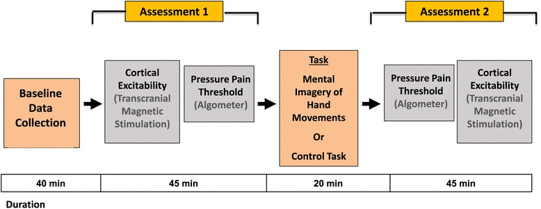
Background: Mental imagery is a powerful method of altering brain activity and behavioral outcomes, such as performance of cognition and motor skills. Further, attention and distraction can modulate pain-related neuronal networks and the perception of pain. This exploratory study examined the effects of mental imagery-induced attention on pressure pain threshold and cortical plasticity using transcranial magnetic stimulation (TMS). This blinded, randomized, and parallel-design trial comprised 30 healthy right-handed male subjects. Exploratory statistical analyses were performed using ANOVA and t-tests for pain and TMS assessments. Pearson's correlation was used to analyze the association between changes in pain threshold and cortical excitability.

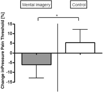
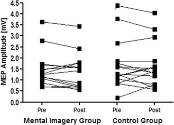
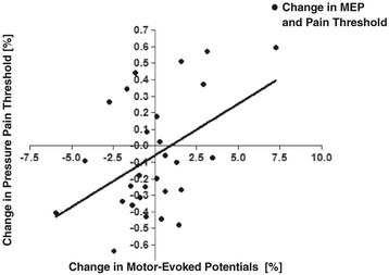
Results: In the analysis of pain outcomes, there was no significant interaction effect on pain between group versus time. In an exploratory analysis, we only observed a significant effect of group for the targeted left hand (ANOVA with pain threshold as the dependent variable and time and group as independent variables). Although there was only a within-group effect of mental imagery on pain, further analyses showed a significant positive correlation of changes in pain threshold and cortical excitability (motor-evoked potentials via TMS).

Conclusions: Mental imagery has a minor effect on pain modulation in healthy subjects. Its effects appear to differ compared with chronic pain, leading to a small decrease in pain threshold. Assessments of cortical excitability confirmed that these effects are related to the modulation of pain-related cortical circuits. These exploratory findings suggest that neuronal plasticity is influenced by pain and that the mental imagery effects on pain depend on the state of central sensitization.


Figures

Figure 1
Figure 1
Relationship between pain perception and motor cortex excitability. a: Relationship and pathway of pain perception and motor cortex excitability. b: Transcranial magnetic stimulation (TMS) and transcranial direct current stimulation (tDCS) can increase motor cortical excitability and pain threshold (= > decrease in pain perception). c: Motor tasks and motor observation increase pain threshold and motor excitability. d: Mental imagery decreases pain threshold and motor cortical excitability. Pictures modified from Flor [8]; Fregni et al. [9]; and Volz et al. [10].
Figure 2
Figure 2
Pain threshold. Pain threshold levels for mental imagery and control groups. Ordinate: Changes in pressure pain threshold level as percentage with respect to baseline value (expressed as: [(t2-t1)/t1] ×100). *p < 0.05 as tested with ANOVA (F(1,27) = 7.40, p = 0.0079) with pressure pain threshold as the dependent variable and group (mental imagery vs control) and time (pre- vs post-intervention) as independent variables. Note that the interaction analyses did not reveal significant results.
Figure 3
Figure 3
Motor-evoked potentials. MEP amplitudes in mV of both groups pre- and postintervention.
Figure 4
Figure 4
Correlation of changes in pain thresholds and MEP amplitude. Correlation between pain threshold of the left hand and MEP amplitudes. Ordinate: Change in pressure pain threshold [in %]. Abscissa: Change of MEP amplitude [in %]; (r = 0.46; p = 0.015). Changes in pressure pain threshold were calculated as follows: t2 (postintervention value) – t1 (preintervention value). Changes in MEP amplitude were calculated as follows: t2 (postintervention amplitude) – t1 (preintervention amplitude).
Figure 5
Figure 5
Study design. Study design and duration of experiment.

References

    1. Pearson DG, Deeprose C, Wallace-Hadrill SM, Burnett Heyes S, Holmes EA. Assessing mental imagery in clinical psychology: a review of imagery measures and a guiding framework. Clin Psychol Rev. 2012;33:1–23. doi: 10.1016/j.cpr.2012.09.001. - DOI - PMC - PubMed
    1. Schuster C, Butler J, Andrews B, Kischka U, Ettlin T. Comparison of embedded and added motor imagery training in patients after stroke: study protocol of a randomised controlled pilot trial using a mixed methods approach. Trials. 2009;10:97. doi: 10.1186/1745-6215-10-97. - DOI - PMC - PubMed
    1. Malouin FRC, Doyon J, Desrosiers J, Belleville S. Training mobility tasks after stroke with combined mental and physical practice: a feasibility study. Neurorehabil Neural Repair. 2004;18:66–75. doi: 10.1177/0888439004266304. - DOI - PubMed
    1. Hoyek N, Di Rienzo F, Collet C, Hoyek F, Guillot A. The therapeutic role of motor imagery on the functional rehabilitation of a stage II shoulder impingement syndrome. Disabil Rehabil. 2014;36(13):1113–9. doi: 10.3109/09638288.2013.833309. - DOI - PubMed
    1. Hussey EP, Smolinsky JG, Piryatinsky I, Budson AE, Ally BA. Using mental imagery to improve memory in patients with Alzheimer disease: trouble generating or remembering the mind’s eye? Alzheimer Dis Assoc Disord. 2011;26:124–34. doi: 10.1097/WAD.0b013e31822e0f73. - DOI - PMC - PubMed

Publication types