

Developmental sex differences in resting state functional connectivity of amygdala sub-regions
- PMID: 25887261
- PMCID: PMC4461484
- DOI: 10.1016/j.neuroimage.2015.04.013
Developmental sex differences in resting state functional connectivity of amygdala sub-regions
Abstract
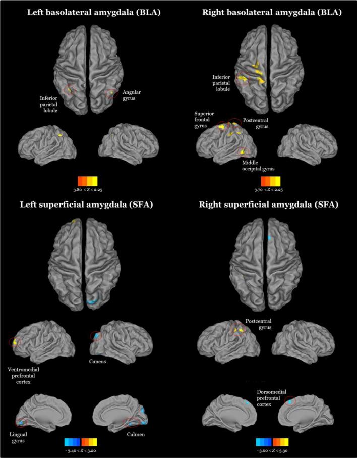
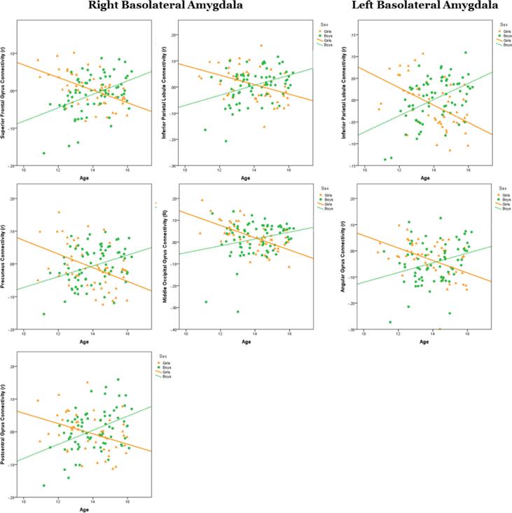
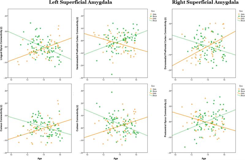
During adolescence, considerable social and biological changes occur that interact with functional brain maturation, some of which are sex-specific. The amygdala is one brain area that has displayed sexual dimorphism, specifically in socio-affective (superficial amygdala [SFA]), stress (centromedial amygdala [CMA]), and learning and memory (basolateral amygdala [BLA]) processing. The amygdala has also been implicated in mood and anxiety disorders which display sex-specific features, most prominently observed during adolescence. Using functional magnetic resonance imaging (fMRI), the present study examined the interaction of age and sex on resting state functional connectivity (RSFC) of amygdala sub-regions, BLA and SFA, in a sample of healthy adolescents between the ages 10 and 16 years (n = 122, 71 boys). Whole-brain, voxel-wise partial correlation analyses were conducted to determine RSFC of bilateral BLA and SFA seed regions, created using the Eickhoff-Zilles maximum probability maps based on cytoarchitectonic mapping and FMRIB's Integrated Registration and Segmentation Tool (FIRST). Monte Carlo simulation was implemented to correct for multiple comparisons (threshold of 53 contiguous voxels with a z-value ≥ 2.25). Results indicated that with increasing age, there was a corresponding decrease in RSFC between both amygdala sub-regions and parieto-occipital cortices, with a concurrent increase in RSFC with medial prefrontal cortex (mPFC). Specifically, boys and girls demonstrated increased coupling of mPFC and left and right SFA with age, respectively; however, neither sex showed increased connectivity between mPFC and BLA, which could indicate relative immaturity of fronto-limbic networks that is similar across sex. A dissociation in connectivity between BLA- and SFA-parieto-occipital RSFC emerged, in which girls had weaker negative RSFC between SFA and parieto-occipital regions and boys had weaker negative RSFC of BLA and parieto-occipital regions with increased age, both standing in contrast to adult patterns of amygdala sub-regional RSFC. The present findings suggest relative immaturity of amygdala sub-regional RSFC with parieto-occipital cortices during adolescence, with unique patterns in both sexes that may support memory and socio-affective processing in boys and girls, respectively. Understanding the underlying normative functional architecture of brain networks associated with the amygdala during adolescence may better inform future research of the neural features associated with increased risk for internalizing psychopathology.
Keywords: Adolescence; Amygdala; Resting state functional connectivity; Sex differences.
Copyright © 2015 Elsevier Inc. All rights reserved.
Figures
References
-
- Amodio DM, Frith CD. Meeting of minds: the medial frontal cortex and social cognition. Nature reviews Neuroscience. 2006;7:268–277. - PubMed
-
- Amunts K, Kedo O, Kindler M, Pieperhoff P, Mohlberg H, Shah NJ, Habel U, Schneider F, Zilles K. Cytoarchitectonic mapping of the human amygdala, hippocampal region and entorhinal cortex: intersubject variability and probability maps. Anatomy and embryology. 2005;210:343–352. - PubMed
-
- Anand A, Li Y, Wang Y, Wu J, Gao S, Bukhari L, Mathews VP, Kalnin A, Lowe MJ. Activity and connectivity of brain mood regulating circuit in depression: a functional magnetic resonance study. Biological psychiatry. 2005;57:1079–1088. - PubMed
Publication types
MeSH terms
Grants and funding
LinkOut - more resources
Full Text Sources
Other Literature Sources
