. 2015 Jun 12;290(24):14893-903.
doi: 10.1074/jbc.M115.652693. Epub 2015 Apr 17.

Tau Trimers Are the Minimal Propagation Unit Spontaneously Internalized to Seed Intracellular Aggregation

Affiliations

Tau Trimers Are the Minimal Propagation Unit Spontaneously Internalized to Seed Intracellular Aggregation

Hilda Mirbaha et al. J Biol Chem. .

Abstract

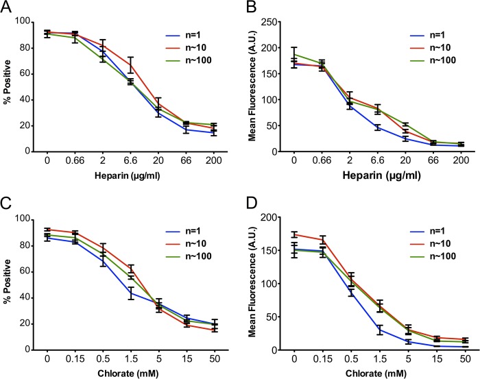
Tau amyloid assemblies propagate aggregation from the outside to the inside of a cell, which may mediate progression of the tauopathies. The critical size of Tau assemblies, or "seeds," responsible for this activity is currently unknown, but this could be important for the design of effective therapies. We studied recombinant Tau repeat domain (RD) and Tau assemblies purified from Alzheimer disease (AD) brain composed largely of full-length Tau. Large RD fibrils were first sonicated to create a range of assembly sizes. We confirmed our ability to resolve stable assemblies ranging from n = 1 to >100 units of Tau using size exclusion chromatography, fluorescence correlation spectroscopy, cross-linking followed by Western blot, and mass spectrometry. All recombinant Tau assemblies bound heparan sulfate proteoglycans on the cell surface, which are required for Tau uptake and seeding, because they were equivalently sensitive to inhibition by heparin and chlorate. However, cells only internalized RD assemblies of n ≥ 3 units. We next analyzed Tau assemblies from AD or control brains. AD brains contained aggregated species, whereas normal brains had predominantly monomer, and no evidence of large assemblies. HEK293 cells and primary neurons spontaneously internalized Tau of n ≥ 3 units from AD brain in a heparin- and chlorate-sensitive manner. Only n ≥ 3-unit assemblies from AD brain spontaneously seeded intracellular Tau aggregation in HEK293 cells. These results indicate that a clear minimum size (n = 3) of Tau seed exists for spontaneous propagation of Tau aggregation from the outside to the inside of a cell, whereas many larger sizes of soluble aggregates trigger uptake and seeding.

Keywords: Tau protein; amyloid; endocytosis; neurodegenerative disease; prion; structure.


Figures

FIGURE 1.
FIGURE 1.
Isolation of recombinant Tau RD fibrillar assemblies by SEC. A, recombinant Tau RD fibrils labeled with AF647 were sonicated for different periods of time (10, 50, and 100 min) to create assemblies in a range of sizes. Assemblies were resolved by SEC with a Sephacryl S500 column. B, the size of each fraction's content was estimated based on the molecular weight of gel filtration standards. C, selected chromatography fractions were resolved by SDS-PAGE probed with a Tau polyclonal antibody. The left lane shows recombinant RD fibril prior to fractionation. Fractions are indicated in the table, as are estimated Tau assembly sizes (where n represents the putative number of Tau units). D, fluorescence of fractions correlates well with protein content of each using a Micro BCA assay. E, selected fractions cross-linked with PFA were resolved by SDS-PAGE and probed with a Tau polyclonal antibody. F, mass spectrometry analysis of cross-linked monomer fraction indicates a molecular mass of 15,376 Da. Analysis of cross-linked trimer fraction indicates three detected species of 45,716 Da (trimer), 30,751 Da (dimer), and 15,376 Da (monomer). H, stability of Tau assemblies was tested by rerunning three fractions (monomer, trimer, and ∼40-mer) separately through the SEC column. Each assembly was stable through the isolation protocol. I, two fractions were recombined (trimer and ∼40-mer) and reisolated using SEC. There was no evidence of interassembly interaction.
FIGURE 2.
FIGURE 2.
Size estimation of RD-647 assemblies by fluorescence correlation spectroscopy. A–F, diffusion time of Tau RD species from monomer through >40-mer. Assemblies labeled with AF647 were measured by FCS to evaluate the accuracy of SEC in identifying the sizes of Tau RD oligomers. Measurements were repeated 10 times each for 30 s. Theoretical autocorrelation functions for single components are indicated as dark lines, with the actual data indicated in red. The residuals of the fit are indicated below each autocorrelation curve.
FIGURE 3.
FIGURE 3.
Tau assemblies bind to the cell surface. A and B, flow cytometry was used to quantify binding of labeled Tau RD assemblies to the plasma membrane. HEK293 cells were treated with AF647-containing buffer (indicated as 0 in the graph) or a 100 nm concentration (monomer equivalent) of selected Tau fractions at 4 °C for 1 h, which allows surface binding but not uptake. Tau units indicate the estimated size of each assembly. There was no difference in the percentage of cells binding any Tau assemblies (A) or the mean fluorescence of each positive cell (B). C and D, Tau RD in selected fractions (n = 1, 10, and 100) was titrated for the cell binding assay. Saturation occurred at approximately the same concentration of Tau (monomer equivalent) for each fraction, although this represents smaller numbers of particles for multimers. A total of 10,000 cells were analyzed for each condition, run in triplicate. Error bars, S.E. of three independent experiments.
FIGURE 4.
FIGURE 4.
Tau assemblies similarly bind HSPGs. Heparin and sodium chlorate were used to inhibit of Tau/HSPG interactions. Heparin binds Tau and prevents its interaction with heparan moieties of HSPGs. Chlorate interferes with HSPG sulfation. HEK293 cells were treated with 100 nm recombinant RD assemblies (n = 1, 10, 100) in the presence of inhibitors. A and B, Tau fractions were incubated with different concentrations of heparin overnight prior to the addition to cells. 10,000 cells were counted per condition in triplicate by flow cytometry. There was no difference in sensitivity to heparin across different assemblies. C and D, cells were treated for 24 h with chlorate at the indicated concentrations prior to exposure to different Tau assemblies. There was no significant difference in sensitivity to chlorate. Error bars, S.E.
FIGURE 5.
FIGURE 5.
Tau RD trimer is the minimal size for uptake. A and B, HEK293 cells were treated for 3 h at 37 °C with a 50 nm concentration (monomer equivalent) of the indicated Tau RD assemblies labeled with AF647 prior to trypsin treatment and quantification of uptake by flow cytometry. Tau assemblies of n = 3 were taken up into cells less efficiently than slightly larger assemblies but more efficiently than assemblies of n = 1 or 2, which were not above background. This could be observed by the percentage of positive cells (A) and the mean fluorescence intensity of positive cells (B). MFI, mean fluorescence intensity; error bars, S.E.
FIGURE 6.
FIGURE 6.
A split-luciferase assay determines that trimers are the minimal seed. A, click beetle luciferase split into two halves (NLuc and CLuc) can dimerize to create a functional holoenzyme. Tau RD(P301S) was fused either to the amino- or carboxyl-terminal halves of luciferase (termed RD-NLuc and Tau-CLuc, respectively). These were stably expressed in HEK293 cells. Upon induced aggregation by the addition of exogenous Tau seeds, luciferase activity is created. B, HEK293 cells stably expressing RD-Cluc/Nluc were exposed in quadruplicate to increasing doses of recombinant Tau RD fibrils. Relative luminescence in comparison with untreated cells was determined 24 h later using a plate-based luminometer. *, p ≤ 0.0001 (analysis of variance). C, HEK293 cells stably expressing RD-Nluc/Cluc were treated with 50 nm Tau RD assemblies, and luminescence was determined. *, p ≤ 0.001; **, p ≤ 0.0001. Error bars, S.E. of three independent experiments.
FIGURE 7.
FIGURE 7.
Purification of brain-derived Tau assemblies. Endogenous Tau was immunopurified from brain lysate, followed by conjugation to AF647. Tau was separated by SEC, monitoring the fluorescence of each fraction. A, control brain featured predominantly Tau monomer. B, AD brain homogenate featured a range of assemblies. C and D, comparison of fluorescence with the protein content of each fraction measured by a Micro BCA assay indicated significant correlation for normal brain (C) as well as AD brain (D). E, SDS-PAGE analysis of recombinant full-length (2N, 4R) Tau (FL), repeat domain Tau (RD), and brain-derived Tau (BD). F, Western blotting of selected SEC fractions of AD brain-derived Tau showed that FL Tau predominates in the fractions. Blots were probed with polyclonal anti-Tau antibody.
FIGURE 8.
FIGURE 8.
Brain-derived Tau assemblies of n ≥ 3 feature spontaneous uptake and seeding. A and B, HEK293 cells (green bars) and primary cortical neurons (blue bars) were treated with a 50 nm concentration of selected AF647-labeled Tau assemblies for 3 h at 37 °C before quantification by flow cytometry. Trimers were the smallest assemblies taken up by HEK293 cells and primary cultured neurons. C, seeding efficiency of Tau was evaluated by a split-luciferase complementation assay. Trimers and larger Tau assemblies from AD brain lysate induced intracellular aggregation compared with cells treated with buffer. Tau derived from normal brain (Nl) did not. *, p ≤ 0.0001 compared with cells treated with buffer. D, selected fractions were incubated with three doses of heparin overnight. Heparin inhibited seeding of all three fractions (n = 3, ∼10, and ∼20) dose-dependently (green). Preincubation of cells with sodium chlorate overnight inhibited seeding dose-dependently (blue). The yellow column on the left indicates background luminescence; red columns indicate vehicle control. *, p value ≤ 0.05; **, p value ≤ 0.0001, compared with untreated sample. Error bars, S.E. from three independent experiments using three different AD brain lysates, each assayed in technical quadruplicate.

References

    1. Lee V. M.-Y., Goedert M., Trojanowski J. Q. (2001) Neurodegenerative tauopathies. Annu. Rev. Neurosci. 24, 1121–1159 - PubMed
    1. Clavaguera F., Bolmont T., Crowther R. A., Abramowski D., Frank S., Probst A., Fraser G., Stalder A. K., Beibel M., Staufenbiel M., Jucker M., Goedert M., Tolnay M. (2009) Transmission and spreading of tauopathy in transgenic mouse brain. Nat. Cell Biol. 11, 909–913 - PMC - PubMed
    1. Clavaguera F., Lavenir I., Falcon B., Frank S., Goedert M., Tolnay M. (2013) “Prion-like” templated misfolding in tauopathies. Brain Pathol. 23, 342–349 - PMC - PubMed
    1. de Calignon A. A., Polydoro M. M., Suárez-Calvet M., William C., Adamowicz D. H. D., Kopeikina K. J. K., Pitstick R., Sahara N., Ashe K. H. K., Carlson G. A., Spires-Jones T. L. T., Hyman B. T. (2012) Propagation of tau pathology in a model of early Alzheimer's disease. Neuron 73, 685–697 - PMC - PubMed
    1. Frost B., Jacks R. L., Diamond M. I. (2009) Propagation of Tau misfolding from the outside to the inside of a cell. J. Biol. Chem. 284, 12845–12852 - PMC - PubMed

Publication types

LinkOut - more resources