. 2015 Feb 26;19(1):53.
doi: 10.1186/s13054-015-0782-3.

Neutralization of osteopontin attenuates neutrophil migration in sepsis-induced acute lung injury

Affiliations

Neutralization of osteopontin attenuates neutrophil migration in sepsis-induced acute lung injury

Yohei Hirano et al. Crit Care. .

Abstract

Introduction: Sepsis refers to severe systemic inflammation leading to acute lung injury (ALI) and death. Introducing novel therapies can reduce the mortality in ALI. Osteopontin (OPN), a secretory glycoprotein produced by immune reactive cells, plays a deleterious role in various inflammatory diseases. However, its role in ALI caused by sepsis remains unexplored. We hypothesize that treatment with an OPN-neutralizing antibody (anti-OPN Ab) protects mice against ALI during sepsis.

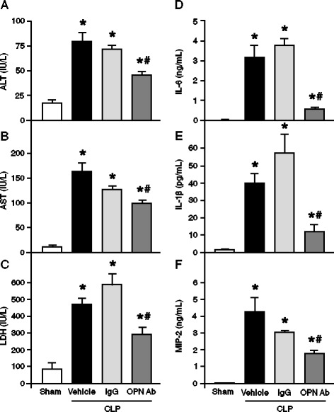
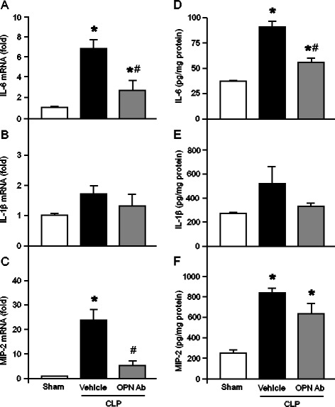
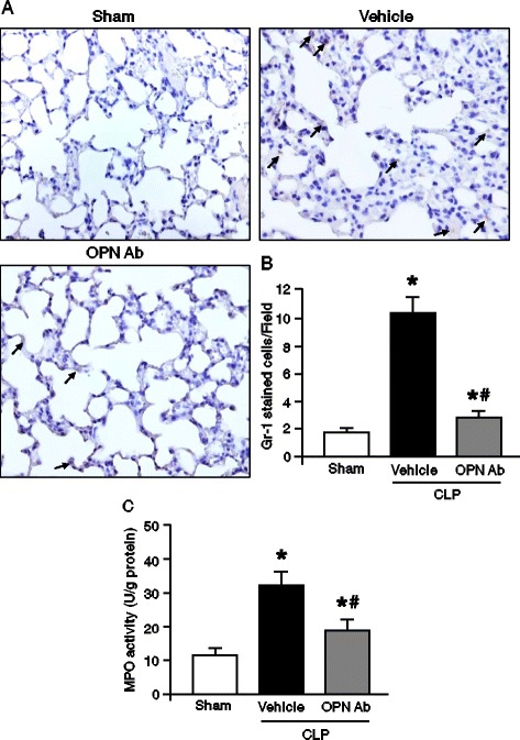
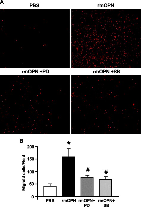
Methods: Sepsis was induced in 8-week-old male C57BL/6 mice by cecal ligation and puncture (CLP). Anti-OPN Ab or non-immunized IgG as control, at a dose of 50 μg/mouse, was intravenously injected at the time of CLP. After 20 hours, the expression of OPN and proinflammatory cytokines in tissues and plasma was examined by real-time PCR, Western blot, and ELISA. Plasma levels of alanine aminotransferase (ALT), aspartate aminotransferase (AST) and lactate dehydrogenase (LDH) and the lung myeloperoxidase (MPO) levels were determined by colorimetric assays. Lung damage and neutrophil infiltrations were determined by histological H&E and Gr-1 staining, respectively. The effect of recombinant mouse OPN (rmOPN) on human neutrophil-like cell (HL-60) migration was performed by Boyden chamber assays and the involvement of intracellular signaling molecules in HL-60 cells was revealed by Western blot.

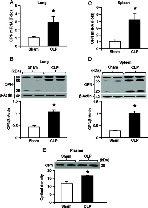
Results: After 20 hours of sepsis, mRNA and protein levels of OPN were significantly induced in lungs, spleen, and plasma. Treatment with an anti-OPN Ab in septic mice significantly reduced the plasma levels of ALT, AST, and LDH, and the proinflammatory cytokines IL-6, IL-1β and the chemokine MIP-2, compared with the vehicle group. Similarly, the lung mRNA and protein expressions of proinflammatory cytokines and chemokine were greatly reduced in anti-OPN Ab-treated animals. The lung histological architecture, MPO and neutrophil infiltration were significantly improved in anti-OPN Ab-treated mice compared with the vehicle animals. Treatment of rmOPN in HL-60 cells significantly increased their migration, in vitro. The neutrophils treated with rmOPN remarkably increased the levels of phospho focal adhesion kinase (pFAK), phospho extracellular signal-regulated kinase (pERK) and phospho p38.

Conclusions: Our findings clearly demonstrate the beneficial outcomes of anti-OPN Ab treatment in protecting against ALI, implicating a novel therapeutic strategy in sepsis.


Figures

Figure 1
Figure 1
Expression of OPN in lungs, spleen and plasma after sepsis in mice . Lungs, spleen and blood samples were harvested at 20 h after CLP or sham operation. Lung expression of OPN at its (A) RNA and (B) protein levels was measured by using real-time PCR and Western blot, respectively. OPN expression in the spleen tissues was determined at its (C) RNA and (D) protein levels. OPN expression in each sample was normalized to β-actin expression and the value of sham group was designated as one for comparison. (E) OPN expression in 3.0 μL of plasma from sham and CLP animals was determined by Western blot. Each blot was quantified by densitometry analysis. Representative blots against OPN and β-actin are shown. Data are expressed as means ± SEM (n = 5 mice/group) and compared by Student’s t test (* P <0.05 vs. shams). CLP, cecal ligation and puncture; OPN, osteopontin; PCR, polymerase chain reaction; SEM, standard error of the mean.
Figure 2
Figure 2
Effect of anti-OPN Ab treatment on plasma levels of organ injury markers and proinflammatory cytokines and chemokine in CLP animals . Sepsis was induced in mice by CLP and anti-OPN Ab or non-immunized IgG control at a dose of 50 μg/mice in 100 μl volumes was injected through the jugular vein. In the vehicle group, 100 μl of PBS was injected in CLP mice via the jugular vein. Blood samples were drawn by cardiac puncture at 20 h of sham-operated, vehicle and anti-OPN Ab-treated mice for measuring (A) ALT, (B) AST and (C) LDH. Similarly, the blood samples collected at 20 h after CLP were measured for (D) IL-6, (E) IL-1β and (F) MIP-2 by ELISA. Data are expressed as means ± SEM (n = 5 mice/group) and compared by one-way ANOVA and SNK method (* P <0.05 vs. shams; # P <0.05 vs. vehicle). Ab, antibody; ALT, alanine aminotransferase; ANOVA, analysis of variance; AST, aspartate aminotransferase; CLP, cecal ligation and puncture; ELISA, enzyme-linked immunosorbent assay; IgG, immunoglobulin G; IL, interleukin; LDH, lactate dehydrogenase; MIP-2, macrophage inflammatory protein 2; OPN, osteopontin; PBS, phosphate-buffered saline; SEM, standard error of the mean; SNK, Student-Newman-Keuls.
Figure 3
Figure 3
Effect of anti-OPN Ab on the expression of proinflammatory cytokines and chemokine in the lungs after CLP . Mice were subjected to CLP and 100 μl of anti-OPN Ab (50 μg/mouse) or PBS as vehicle was injected through the jugular vein. Lung tissues were collected after 20 h from sham-operated, vehicle, and anti-OPN Ab-treated mice. The tissue expression of (A) IL-6, (B) IL-1β and (C) MIP-2 was determined by real-time PCR. Gene expression was normalized to β-actin. The sham expression level was designated as one for comparison. Similarly, 50 μg of total protein extracted from the lung tissues were examined for (D) IL-6, (E) IL-1β and (F) MIP-2 analysis by ELISA. Finally, the results are expressed as per mg of proteins. Data are represented as means ± SEM (n = 5 mice/group) and compared by one-way ANOVA and SNK method (* P <0.05 vs. sham; # P <0.05 vs. vehicle). Ab, antibody; ANOVA, analysis of variance; CLP, cecal ligation and puncture; ELISA, enzyme-linked immunosorbent assay; IL, interleukin; MIP-2, macrophage inflammatory protein 2; OPN, osteopontin; PBS, phosphate-buffered saline; PCR, polymerase chain reaction; SEM, standard error of the mean; SNK, Student-Newman-Keuls.
Figure 4
Figure 4
Evaluation of lung histology in mice after CLP . Lung tissues were harvested after 20 h from sham-operated, vehicle and anti-OPN Ab-treated mice and stained with H&E. Slides were observed under light microscopy at × 200 original magnifications (inset: ×400 original magnification). (A) Representative images for sham, vehicle, and anti-OPN Ab treatment groups are shown. (B) Histological injury scores of the lungs in different groups were quantified as described in the Materials and Methods. Data are expressed as means ± SEM (n = 5 mice/group) and compared by one-way ANOVA and SNK method (* P <0.05 vs. shams; # P <0.05 vs. vehicles). Ab, antibody; ANOVA, analysis of variance; CLP, cecal ligation and puncture; H&E, hematoxylin and eosin; OPN, osteopontin; SEM, standard error of the mean; SNK, Student-Newman-Keuls.
Figure 5
Figure 5
Assessment of neutrophil infiltration into the lungs after CLP . Lung tissues were collected at 20 h after sham-operation, vehicle and anti-OPN Ab treatment in mice. (A) Representative images of the lung tissue sections with immunostaining against Gr-1 at × 200 original magnification are shown. Arrows demarcate examples of areas of staining Gr-1-positive cells. (B) A graphical representation of Gr-1-positive cells averaged over 10 microscopic fields per animal lung tissues. (C) Myeloperoxidase (MPO) activities in lung tissues were determined spectrophotometrically. Data are expressed as means ± SEM (n = 5 mice/group) and compared by one-way ANOVA and SNK method (* P <0.05 vs. shams; # P <0.05 vs. vehicles). Ab, antibody; ANOVA, analysis of variance; CLP, cecal ligation and puncture; OPN, osteopontin; SEM, standard error of the mean; SNK, Student-Newman-Keuls.
Figure 6
Figure 6
rmOPN-mediated neutrophil migration in vivo and in vitro. C57BL/6 mice were injected with rmOPN at a dose of 2.5 μg/mice, intratracheally. After 20 h, cells from the lung tissues were isolated and stained with APC-anti-Gr-1 Ab and then subjected to flow cytometry. (A) Representative dot blots indicating the percentages of Gr-1-positive cells are shown. (B) The mean percentages of Gr-1-positive cells obtained from PBS- and rmOPN-injected mice are shown. Data are expressed as means ± SEM (n = 4 mice/group) and compared by Student’s t test (* P <0.05 vs. PBS). (C) A total of 5 × 105 primary neutrophil cells isolated from mouse bone marrow were placed into the insert of a Boyden chamber. The bottom compartment contained the RPMI medium with PBS or rmOPN at a dose of 10 μg/ml as a chemotactic stimulus. After 2 h, the migrated primary neutrophil cells were counted. A representative image of the migrated primary neutrophil cells labeled with PI (red fluorescence) on the bottom of the transwell membrane is shown. Cells were observed at × 200 original magnification. (B) Migrated primary neutrophils were counted in five random microscopic fields per well and averaged in each group. Data are expressed as means ± SEM (n = 4/group) and compared by Student’s t test (* P <0.05 vs. PBS). Ab, antibody; OPN, osteopontin; PBS, phosphate-buffered saline; PI, propidium iodide; rmOPN, recombinant mouse OPN; SEM, standard error of the mean.
Figure 7
Figure 7
rmOPN-mediated activation of FAK and MAP kinase signaling molecules in dHL-60 cells . Differentiated HL-60 cells were incubated with PBS or rmOPN at a dose of 2 μg/mL for 90 min. (A) The status of phosphorylated FAK (pFAK) and β-actin, (B) pERK and total ERK and (C) pp38 and total p38 in each group was determined by Western blot. Blots were scanned and quantified with densitometry. Representative blots against these proteins are shown. Data are expressed as means ± SEM obtained from two independent experiments (n = 5/group) and compared by Students t test (*P <0.05 vs. PBS). ERK, extracellular signal-regulated protein kinase; FAK, focal adhesion kinase; MAP, mitogen-activated protein; OPN, osteopontin; PBS, phosphate-buffered saline; rmOPN, recombinant mouse OPN; SEM, standard error of the mean.
Figure 8
Figure 8
Effect of rmOPN on dHL-60 cell migration in vitro. Boyden chamber assay was performed as described in Materials and Methods. (A) A representative image of migrated dHL-60 cells labeled with PI (red fluorescence) on the bottom of the transwell membrane. Cells were observed at × 200 original magnification. (B) Migrated dHL-60 cells were counted in five random microscopic fields per well and averaged in each group. Data are expressed as means ± SEM (n = 4/group) and compared by one-way ANOVA and SNK method (* P <0.05 vs. PBS; # P <0.05 vs. rmOPN). ANOVA, analysis of variance; OPN, osteopontin; PBS, phosphate-buffered saline; PI, propidium iodide; rmOPN, recombinant mouse OPN; SEM, standard error of the mean; SNK, Student-Newman-Keuls.

References

    1. Aziz M, Jacob A, Yang WL, Matsuda A, Wang P. Current trends in inflammatory and immunomodulatory mediators in sepsis. J Leukoc Biol. 2013;93:329–42. doi: 10.1189/jlb.0912437. - DOI - PMC - PubMed
    1. Rangel-Frausto MS, Pittet D, Costigan M, Hwang T, Davis CS, Wenzel RP. The natural history of the systemic inflammatory response syndrome (SIRS): a prospective study. JAMA. 1995;273:117–23. doi: 10.1001/jama.1995.03520260039030. - DOI - PubMed
    1. Angus DC, Linde-Zwirble WT, Lidicker J, Clermont G, Carcillo J, Pinsky MR. Epidemiology of severe sepsis in the United States: analysis of incidence, outcome, and associated costs of care. Crit Care Med. 2001;29:1303–10. doi: 10.1097/00003246-200107000-00002. - DOI - PubMed
    1. Matuschak GM, Lechner AJ. Acute lung injury and the acute respiratory distress syndrome: pathophysiology and treatment. Mo Med. 2010;107:252–8. - PMC - PubMed
    1. Rubenfeld GD, Caldwell E, Peabody E, Weaver J, Martin DP, Neff M, et al. Incidence and outcomes of acute lung injury. N Engl J Med. 2005;353:1685–93. doi: 10.1056/NEJMoa050333. - DOI - PubMed

Publication types