. 2015 Feb 27;19(1):59.
doi: 10.1186/s13054-015-0785-0.

Endotracheal tube biofilm translocation in the lateral Trendelenburg position

Affiliations

Endotracheal tube biofilm translocation in the lateral Trendelenburg position

Gianluigi Li Bassi et al. Crit Care. .

Abstract

Introduction: Laboratory studies demonstrated that the lateral Trendelenburg position (LTP) is superior to the semirecumbent position (SRP) in the prevention of ventilator-associated pulmonary infections. We assessed whether the LTP could also prevent pulmonary colonization and infections caused by an endotracheal tube (ETT) biofilm.

Methods: Eighteen pigs were intubated with ETTs colonized by Pseudomonas aeruginosa biofilm. Pigs were positioned in LTP and randomized to be on mechanical ventilatin (MV) up to 24 hour, 48 hour, 48 hour with acute lung injury (ALI) by oleic acid and 72 hour. Bacteriologic and microscopy studies confirmed presence of biofilm within the ETT. Upon autopsy, samples from the proximal and distal airways were excised for P.aeruginosa quantification. Ventilator-associated tracheobronchitis (VAT) was confirmed by bronchial tissue culture ≥3 log colony forming units per gram (cfu/g). In pulmonary lobes with gross findings of pneumonia, ventilator-associated pneumonia (VAP) was confirmed by lung tissue culture ≥3 log cfu/g.

Results: P.aeruginosa colonized the internal lumen of 16 out of 18 ETTs (88.89%), and a mature biofilm was consistently present. P.aeruginosa colonization did not differ among groups, and was found in 23.6% of samples from the proximal airways, and in 7.1% from the distal bronchi (P = 0.001). Animals of the 24 hour group never developed respiratory infections, whereas 20%, 60% and 25% of the animals in group 48 hour, 48 hour-ALI and 72 hour developed P.aeruginosa VAT, respectively (P = 0.327). Nevertheless, VAP never developed.

Conclusions: Our findings imply that during the course of invasive MV up to 72 hour, an ETT P.aeruginosa biofilm hastily colonizes the respiratory tract. Yet, the LTP compartmentalizes colonization and infection within the proximal airways and VAP never develops.


Figures

Figure 1
Figure 1
Bronchopulmonary anatomy of the pig lung. The numbers from 1 to 11 indicate sites from which samples were taken for microbiological studies: 1 to 2, trachea; 3 to 4, main bronchi; 5 to 11, segmental bronchi. RUL, right upper lobe; RML, right medium lobe; RLL, right lower lobe; LUL, left upper lobe; LLL, left lower lobe.
Figure 2
Figure 2
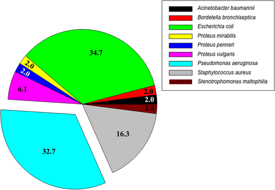
Endotracheal tube colonization. The pie chart represents the bacterial diversity recovered from the endotracheal tube upon extubation. The percentage of Pseudomonas aeruginosa isolates is explicitly indicated.
Figure 3
Figure 3
Confocal laser scanning micrograph of the internal surface of endotracheal tubes of animals mechanically ventilated for 24, 48, 48 hours with concomitant lung injury and 72 hours. The samples were stained with BacLight Live/Dead (magnification × 250). The white arrow indicates the endotracheal tube internal surface. Note in all pictures a fully mature biofilm adherent to the endotracheal tube.
Figure 4
Figure 4
Scanning electron micrographs of the internal surface of the endotracheal tube (ETT) in animals mechanically ventilated for 24, 48, 48 hours with concomitant lung injury and 72 hours. Note in all pictures consistent presence of stage III/IV biofilm, characterized by multiple rod-shaped bacteria embedded within an extracellular polymeric substance. PIG 50, frontal view of the ETT lumen (magnification × 3,000); PIG 30, cross-section of the ETT lumen (magnification × 1,500); PIG 51, frontal view of the ETT lumen (magnification × 3,000); PIG 44, frontal-view of the ETT lumen (magnification × 3,000); PIG 40, frontal view of the ETT lumen (magnification × 3,000); PIG 37, frontal view of the ETT lumen (magnification × 3,000); PIG 48, frontal view of the ETT lumen (magnification × 3,000); PIG 31, frontal view of the ETT lumen (magnification × 3,000).
Figure 5
Figure 5
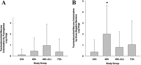
Tracheobronchial mucosa bacterial concentrations for P.aeruginos. (A) and all aerobic Gram-negative bacteria (B) per study group. The mean P.aeruginosa concentration did not differ among groups (P = 0.059), whereas, the concentration of all aerobic Gram-negative bacteria (B) was significantly different (P = 0.002). *P = 0.005 versus group 24 h. 24 h, 24 hours; 48 h, 48 hours; 48 h-ALI, 48 hours with concomitant acute lung injury; 72 h, 72 hours.

References

    1. Torres A, Serra-Batlles J, Ros E, Piera C, Puig de la Bellacasa J, Cobos A, et al. Pulmonary aspiration of gastric contents in patients receiving mechanical ventilation: the effect of body position. Ann Intern Med. 1992;116:540–543. doi: 10.7326/0003-4819-116-7-540. - DOI - PubMed
    1. Drakulovic MB, Torres A, Bauer TT, Nicolas JM, Nogué S, Ferrer M. Supine body position as a risk factor for nosocomial pneumonia in mechanically ventilated patients: a randomised trial. Lancet. 1999;354:1851–1858. doi: 10.1016/S0140-6736(98)12251-1. - DOI - PubMed
    1. Li Bassi G, Marti JD, Saucedo L, Rigol M, Roca I, Cabanas M, et al. Gravity predominates over ventilatory pattern in the prevention of ventilator-associated pneumonia. Crit Care Med. 2014;42:e620–e627. doi: 10.1097/CCM.0000000000000487. - DOI - PubMed
    1. Panigada M, Berra L, Greco G, Stylianou M, Kolobow T. Bacterial colonization of the respiratory tract following tracheal intubation-effect of gravity: an experimental study. Crit Care Med. 2003;31:729–737. doi: 10.1097/01.CCM.0000049943.01252.E5. - DOI - PubMed
    1. Zanella A, Cressoni M, Epp M, Hoffmann V, Stylianou M, Kolobow T. Effects of tracheal orientation on development of ventilator-associated pneumonia: an experimental study. Intensive Care Med. 2012;38:677–685. doi: 10.1007/s00134-012-2495-2. - DOI - PubMed

Publication types