

Crosstalk in glomerular injury and repair
- PMID: 25887901
- PMCID: PMC4465999
- DOI: 10.1097/MNH.0000000000000117
Crosstalk in glomerular injury and repair
Abstract
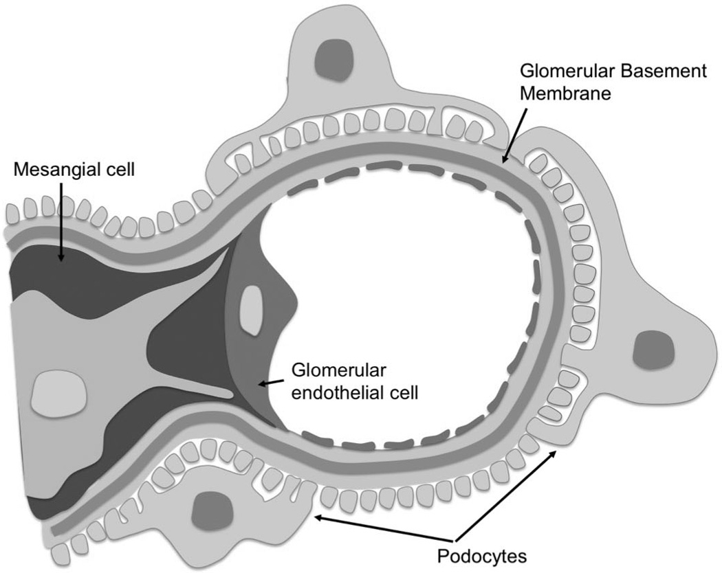
Purpose of review: The glomerulus is a unique structure required for filtration of blood, while retaining plasma proteins based on size and charge selectivity. Distinct cell types form the structural unit that creates the filtration barrier. Structurally, fenestrated endothelial cells line the capillary loops and lie in close contact with mesangial cells. Podocytes are connected by specialized intercellular junctions known as slit diaphragms and separated from the endothelial compartment by the glomerular basement membrane. In order for this highly specialized structure to function, cross-communication between these cells must occur.
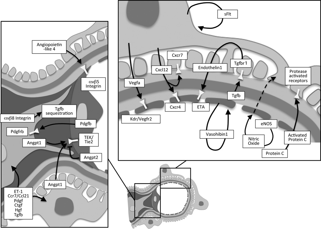
Recent findings: Although classical studies have established key roles for vascular endothelial and platelet-derived growth factors in glomerular cross-communication, novel paracrine signaling pathways within the glomerulus have recently been identified. In addition, unique cellular pathways of established signaling cascades have been identified that are important for maintaining glomerular barrier function in health and disease.
Summary: Here, we will review our current understanding of the processes of cross-communication between the unique cellular constituents forming the glomerular filtration unit. We will highlight recent findings of cellular crosstalk via signaling pathways that regulate glomerular barrier function in pathophysiological conditions.
Conflict of interest statement
The authors declare no conflicts of interest.
Figures
References
-
- Vaughan MR, Quaggin SE. How do mesangial and endothelial cells form the glomerular tuft? J Am Soc Nephrol. 2008;19:24–33. - PubMed
-
- Maezawa Y, Cina D, Quaggin SE. Glomerular Cell Biology. In: Alpern R, Caplan M, editors. Seldin and Giebisch's The Kidney: Physiology & Pathophysiology. Vol. 1. Moe O: Elsevier; 2012. pp. 721–756.
-
- Carmeliet P, Ferreira V, Breier G, et al. Abnormal blood vessel development and lethality in embryos lacking a single VEGF allele. Nature. 1996;380:435–439. - PubMed
Publication types
MeSH terms
Grants and funding
LinkOut - more resources
Full Text Sources
Other Literature Sources
Medical
Research Materials
