. 2015 Apr 10;19(1):159.
doi: 10.1186/s13054-015-0882-0.

Prolonged mechanical ventilation-induced neuroinflammation affects postoperative memory dysfunction in surgical mice

Affiliations

Prolonged mechanical ventilation-induced neuroinflammation affects postoperative memory dysfunction in surgical mice

Chang Chen et al. Crit Care. .

Abstract

Introduction: Patients undergoing surgery frequently develop neuropsychological disturbances, including cognitive decline or memory impairment, and routine clinical procedures such as mechanical ventilation (MV) may affect acute-phase brain outcome. We aimed to investigate the effect of the prolonged MV on postoperative memory dysfunction in surgical mice.

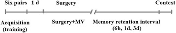
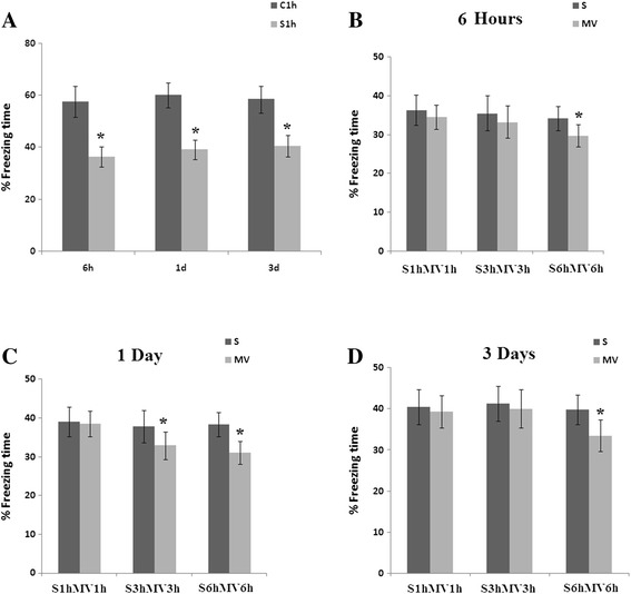
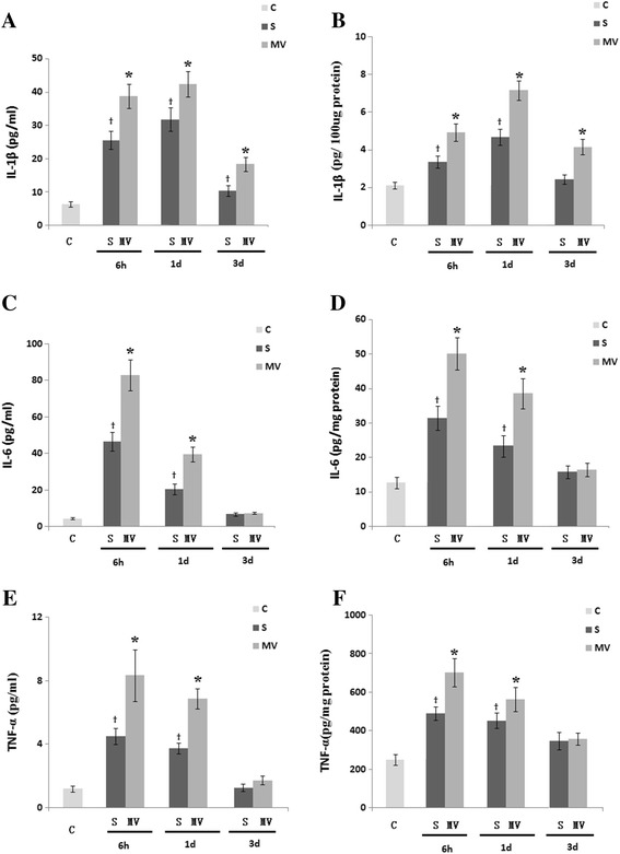
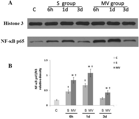
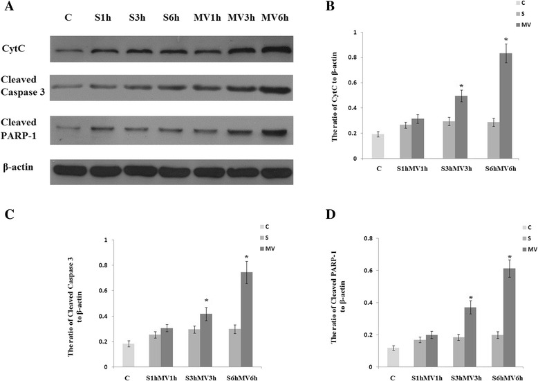
Methods: Male C57BL/6 mice were randomly divided into the following three groups: (1) The control group (group C) comprised anesthetized, unventilated animals; (2) the surgery group (subgroups S1h, S3h and S6h) was unventilated animals that underwent surgery under general anesthesia; and (3) the MV group (subgroups MV1h, MV3h and MV6h) was made up of animals under MV for 1 hour, 3 hours or 6 hours after surgery. Separate cohorts of animals were tested for memory function with fear conditioning tests or were killed at 6 hours, 1 day or 3 days postsurgery or post-MV to examine levels systemic and hippocampal interleukin (IL)-1β, IL-6 and tumor necrosis factor α (TNFα), and assessed synaptic structure and microglial activation. Nuclear factor κB (NF-κB) p65, cytochrome c, cleaved caspase-3 and cleaved poly(ADP-ribose) polymerase (PARP) activation were analyzed by Western blotting.

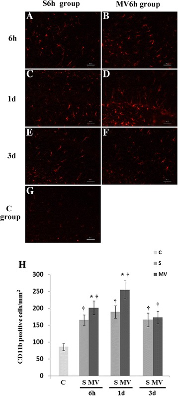
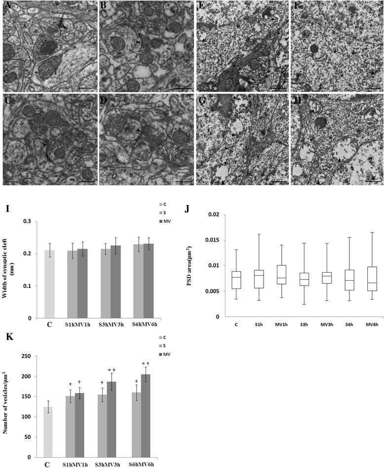
Results: The MV6h group showed increased CD11b-immunopositive cells, synapse degeneration, cytochrome c release, cleaved caspase-3 and cleaved PARP-1 activation after surgery, as well as a decrease in freezing time after surgery. At 6 hours and 1 day post-MV, MV6h increased NF-κB activation and levels of systemic and hippocampal IL-1β, IL-6 and TNFα after surgery.

Conclusions: Prolonged MV after surgery further aggravates cognitive decline that may stem from upregulation of hippocampal IL-1β, IL-6 and TNFα, partially via activation of gliocytes in the surgical mouse hippocampus.


Figures

Figure 1
Figure 1
Assessment of efficiency of consolidation of memory using a contextual fear conditioning protocol. Mice were frightened by an aversive stimulus, in this case tone and electrical foot shock stimulus, to acquire fear memory (acquisition). One day prior to surgery (n =36) or mechanical ventilation (MV; n =36), animals were trained for fear conditioning. At 6 hours, 1 day and 3 days after those treatments, memory was reassessed by measuring the period of time during which the animal became involuntarily immobile when reintroduced to the aversive context.
Figure 2
Figure 2
Contextual fear conditioning responses after surgery followed by mechanical ventilation. In the surgery group (S), surgery consisted of an open tibial fracture with intramedullary fixation in aseptic conditions under general anesthesia with isoflurane and buprenorphine, and then mice were placed in the anesthesia chamber with isoflurane for 1 hour, 3 hours or 6 hours. In the mechanical ventilation (MV) group, after the same surgery, isoflurane was administered to maintain anesthetic level during MV for 1 hour, 3-hour and 6-hour procedures. (A) Freezing time of C1h and S1h subgroups. (B) Percentage freezing time in the 6-hour groups. (C) Percentage freezing time on day 1. (D) Percentage freezing time on day 3. n =12/group. Values are mean ± standard error of the mean. *P <0.05 indicates significant differences.
Figure 3
Figure 3
Effects of mechanical ventilation on the levels of interleukin 1β, interleukin 6 and tumor necrosis factor α after surgery in the plasma and hippocampus. Six-hour exposure to mechanical ventilation (MV) resulted in elevated plasma (A, C and E) and hippocampus (B, D and F) levels of interleukin (IL)-1β, IL-6 and tumor necrosis factor α (TNFα) at both 6 hours and 1 day after MV compared with the surgery group (S), as measured by enzyme-linked immunosorbent assay. Values are mean ± standard error of the mean. n =6/group. *P <0.05, compared with group S; P <0.05 compared with the control group (C).
Figure 4
Figure 4
Six-hour exposure to mechanical ventilation significantly increased nuclear factor κB activation in hippocampal tissues. (A) Nuclear factor κB (NF-κB) p65 expression was detected by Western blot analysis. (B) NF-κB p65 protein expression was significantly increased in the mechanical ventilation (MV) groups compared with the surgery groups (S) at 6 hours, 1 day and 3 days postsurgery or post-MV. Western blot assay data are expressed as mean ± standard error of the mean. n =6/group. *P <0.05 compared with group S. P <0.05 compared with group C.
Figure 5
Figure 5
Immunofluorescence of microglia with anti-CD11b. Hippocampi were harvested 6 hours, 1 days and 3 days postsurgery (S) or after mechanical ventilation (MV). (A) through (G) Representative photomicrographs of tissue from only surgical animals and surgical animals treated with 6 hours of MV (pictures shown refer to CA2 region of the hippocampus in tissue. Scale bars =50 μm. (H) Cell counts reveal significant differences between S6h and MV6h at 6 hours and 1 day postsurgery or post-MV. One day after surgery, mice showed significantly lower levels of reactive microgliosis compared with surgical mice treated with 3 hours or 6 hours of MV. Values are mean ± SEM. n =4/group. *P <0.05 compared with group S; P <0.05 compared with control group (C).
Figure 6
Figure 6
Ultrastructural changes of synapses in the hippocampal CA1 region visualized by transmission electron microscopy. The animals were killed at 6 hours post-MV. Six-hour exposure to mechanical ventilation (MV) aggravated impairment in the synaptic ultrastructure at 6 hours post-MV. (A) Control group (C). (B) Surgical group (S) at 6 hours (S6h). (C) MV group at 3 hours (MV3h). (D) MV group at 6 hours (MV6h). Arrows point to synaptic cleft; arrowheads point to postsynaptic density. Ultrastructural changes of the mitochondrion and endoplasmic reticulum were visualized under transmission electron microscopy. The degree of rough endoplasmic reticulum degranulation in the hippocampus was more severe in the MV6h group (G) and (H) compared with the MV3h group (E) and (F). Scale bar =500 nm. Quantitation of the data is presented for (I) width of synaptic cleft, (J) area of postsynaptic density (PSD) and (K) number of vesicles. Data for each group in (J) are summarized by a box chart, in which the horizontal lines denote the 25th, 50th and 75th percentile values and the error bars denote the 5th and 95th percentile values. *P <0.05, compared with group S; P <0.05, compared with group C.
Figure 7
Figure 7
Mechanical ventilation induced significantly greater apoptosis in surgical mice. Six-hour exposure to mechanical ventilation (MV) induced upregulation of cytochrome c (Cytc), cleaved caspase-3 and cleaved poly(ADP-ribose) polymerase 1 (PARP-1) in the hippocampus after surgery (S). (A) Representative Western blot of Cytc, cleaved caspase-3 and cleaved PARP-1. (B, C and D) The results of semiquantitative analysis of the ratio of Cytc to β-actin (B), the ratio of cleaved caspase-3 to β-actin (C) and the ratio of cleaved PARP-1 to β-actin (D). All Western blot assay data are expressed as mean ± standard error of the mean. C, Control group. n =6/group. *P <0.05, compared with group S.

References

    1. Quílez ME, López-Aguilar J, Blanch L. Organ crosstalk during acute lung injury, acute respiratory distress syndrome, and mechanical ventilation. Curr Opin Crit Care. 2012;18:23–28. doi: 10.1097/MCC.0b013e32834ef3ea. - DOI - PubMed
    1. Brummel NE, Jackson JC, Pandharipande PP, Thompson JL, Shintani AK, Dittus RS, et al. Delirium in the ICU and subsequent long-term disability among survivors of mechanical ventilation. Crit Care Med. 2014;42:369–377. doi: 10.1097/CCM.0b013e3182a645bd. - DOI - PMC - PubMed
    1. Van Rompaey B, Schuurmans MJ, Shortridge-Baggett LM, Truijen S, Elseviers M, Bossaert L. A comparison of the CAM-ICU and the NEECHAM Confusion Scale in intensive care delirium assessment: an observational study in non-intubated patients. Crit Care. 2008;12:R16. doi: 10.1186/cc6790. - DOI - PMC - PubMed
    1. López-Aguilar J, Fernández-Gonzalo MS, Turon M, Quílez ME, Gómez-Simón V, Jódar MM, et al. Lung–brain interaction in the mechanically ventilated patient. Med Intensiva. 2013;37:485–492. doi: 10.1016/j.medin.2012.10.005. - DOI - PubMed
    1. Stevens RD, Puybasset L. The brain–lung–brain axis. Intensive Care Med. 2011;37:1054–1056. doi: 10.1007/s00134-011-2233-1. - DOI - PubMed

Publication types

MeSH terms

Substances