. 2015 Jan 28;19(1):23.
doi: 10.1186/s13054-015-0759-2.

Increasing the inspiratory time and I:E ratio during mechanical ventilation aggravates ventilator-induced lung injury in mice

Affiliations

Increasing the inspiratory time and I:E ratio during mechanical ventilation aggravates ventilator-induced lung injury in mice

Holger C Müller-Redetzky et al. Crit Care. .

Abstract

Introduction: Lung-protective ventilation reduced acute respiratory distress syndrome (ARDS) mortality. To minimize ventilator-induced lung injury (VILI), tidal volume is limited, high plateau pressures are avoided, and positive end-expiratory pressure (PEEP) is applied. However, the impact of specific ventilatory patterns on VILI is not well defined. Increasing inspiratory time and thereby the inspiratory/expiratory ratio (I:E ratio) may improve oxygenation, but may also be harmful as the absolute stress and strain over time increase. We thus hypothesized that increasing inspiratory time and I:E ratio aggravates VILI.

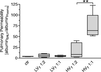
Methods: VILI was induced in mice by high tidal-volume ventilation (HVT 34 ml/kg). Low tidal-volume ventilation (LVT 9 ml/kg) was used in control groups. PEEP was set to 2 cm H2O, FiO2 was 0.5 in all groups. HVT and LVT mice were ventilated with either I:E of 1:2 (LVT 1:2, HVT 1:2) or 1:1 (LVT 1:1, HVT 1:1) for 4 hours or until an alternative end point, defined as mean arterial blood pressure below 40 mm Hg. Dynamic hyperinflation due to the increased I:E ratio was excluded in a separate group of animals. Survival, lung compliance, oxygenation, pulmonary permeability, markers of pulmonary and systemic inflammation (leukocyte differentiation in lung and blood, analyses of pulmonary interleukin-6, interleukin-1β, keratinocyte-derived chemokine, monocyte chemoattractant protein-1), and histopathologic pulmonary changes were analyzed.

Results: LVT 1:2 or LVT 1:1 did not result in VILI, and all individuals survived the ventilation period. HVT 1:2 decreased lung compliance, increased pulmonary neutrophils and cytokine expression, and evoked marked histologic signs of lung injury. All animals survived. HVT 1:1 caused further significant worsening of oxygenation, compliance and increased pulmonary proinflammatory cytokine expression, and pulmonary and blood neutrophils. In the HVT 1:1 group, significant mortality during mechanical ventilation was observed.

Conclusion: According to the "baby lung" concept, mechanical ventilation-associated stress and strain in overinflated regions of ARDS lungs was simulated by using high tidal-volume ventilation. Increase of inspiratory time and I:E ratio significantly aggravated VILI in mice, suggesting an impact of a "stress/strain × time product" for the pathogenesis of VILI. Thus increasing the inspiratory time and I:E ratio should be critically considered.


Figures

Figure 1
Figure 1
Schematic graphic of the respiratory cycle during I:E ratio of 1:2 and 1:1. In all I:E 1:1 groups, the inspiratory time was prolonged by adding an inspiratory hold after completion of lung inflation, thereby leaving pressure and flow acceleration during inspiration identical between the corresponding LVT and HVT 1:2 groups.
Figure 2
Figure 2
Increasing the inspiratory time and I:E ratio during MV increased mortality in VILI. Mice were mechanically ventilated for 4 hours with either low tidal volume (LVT 9 ml/kg) or high tidal volume (HVT 34 ml/kg) and an inspiratory/expiratory ratio of 1:2 or 1:1, respectively. If the mean arterial pressure decreased below 40 mm Hg, the experiment was prematurely terminated, as this predicts death with certainty in this model. n = 13-14 each group; ***P < 0.001.
Figure 3
Figure 3
Increasing the inspiratory time and I:E ratio during MV increased VILI. Mice were mechanically ventilated for 4 hours with either low tidal volume (LVT 9 ml/kg) or high tidal volume (HVT 34 ml/kg) and an inspiratory/expiratory ratio of 1:2 or 1:1, respectively. An alternative end point was defined as decreasing of mean arterial blood pressure below 40 mm Hg, as this predicts death with certainty in this model. Controls (ctr) were subjected to LVT 1:2 ventilation only during operation and were killed before the 4-hour ventilation protocol started. Ctr and LVT groups showed no signs of lung injury macroscopically. HVT 1:2 revealed only subtle macroscopic signs of injury, whereas the HVT 1:1 group showed massive edema formation. Representative images from 13 to 14 animals per group are shown.
Figure 4
Figure 4
Increasing the inspiratory time and I:E ratio during MV increased histopathologic signs of lung injury. Mice were mechanically ventilated for 4 hours with either low tidal volume (LVT 9 ml/kg) or high tidal volume (HVT 34 ml/kg) and an inspiratory/expiratory ratio of 1:2 or 1:1, respectively. An alternative end point was defined as decreasing of mean arterial blood pressure below 40 mm Hg, which predicts death with certainty in this model. Controls (ctr) were subjected to LVT 1:2 ventilation only during operation and were killed before the 4-hour ventilation protocol started. Histopathology of lungs from ctr, HVT 1:2, and HVT 1:1 groups, stained with the periodic acid-Schiff (PAS) reaction, are shown: In contrast to the ctr group, both ventilated groups had damage of the alveolar walls with septal thickening, necrosis, and desquamation of alveolar epithelial cells type I, formation of hyaline membranes (red arrows), and increased numbers of intraalveolar cells (predominantly neutrophils and macrophages, black arrows). PAS reaction highlighted the more severe and more continuous as well as thicker hyaline membranes along the alveolar surfaces of lungs from the HVT 1:1 group. Top panel: magnification × 200; Bottom panel: magnification × 400. Representative images from each group (n = 4 each) are shown.
Figure 5
Figure 5
Increasing the inspiratory time and I:E ratio during MV increased pulmonary permeability in VILI. Mice were mechanically ventilated for 4 hours with either low tidal volume (LVT 9 ml/kg) or high tidal volume (HVT 34 ml/kg) and an inspiratory/expiratory ratio of 1:2 or 1:1, respectively. An alternative end point was defined as decreasing of mean arterial blood pressure below 40 mm Hg, which predicts death with certainty in this model. Controls (ctr) were subjected to LVT 1:2 ventilation only during operation and were killed before the 4-hour ventilation protocol started. Albumin concentrations in bronchoalveolar lavage fluid (BALF) and plasma were determined. An increased albumin BALF/plasma ratio indicated enhanced lung permeability. n = 5 to 6 each group; ** < 0.01.
Figure 6
Figure 6
Increasing the inspiratory time and I:E ratio reduced oxygenation capacity in VILI. Mice were mechanically ventilated for 4 hours with either low tidal volume (LVT 9 ml/kg) or high tidal volume (HVT 34 ml/kg) and an inspiratory/expiratory ratio of 1:2 or 1:1, respectively. An alternative end point was defined as decreasing of mean arterial blood pressure below 40 mm Hg, which predicts death with certainty in this model. Controls (ctr) were subjected to LVT 1:2 ventilation only during operation and were killed before the 4-hour ventilation protocol started. (A) Pulse oximetry revealed stable oxygen saturation in LVT 1:2, LVT 1:1, and HVT 1:2 groups, whereas the HVT 1:1 ventilated mice developed a decrease of oxygen saturation during the 4-hour ventilation period. End-point measurements of arterial partial pressure of oxygen were performed, and the P/F ratio (B), and the oxygenation index were calculated (C). n = 13 to 14 in each group; ***P < 0.001.
Figure 7
Figure 7
Increasing the inspiratory time and I:E ratio deteriorated lung function in VILI. Mice were mechanically ventilated for 4 hours with either low tidal volume (LVT 9 ml/kg) or high tidal volume (HVT 34 ml/kg) and an inspiratory/expiratory ratio of 1:2 or 1:1, respectively. An alternative end point was defined as decreasing of mean arterial blood pressure below 40 mm Hg, which predicts death with certainty in this model. Controls (ctr) were subjected to LVT 1:2 ventilation only during operation and were killed before the 4-hour ventilation protocol started. (A) Dynamic elastance (E) was measured every 10 minutes during the experiment. Results of end-point measurements of dynamic compliance (B) and static compliance (C) are shown. n = 13 to 14 in each group; ***P < 0.001.
Figure 8
Figure 8
Increasing the inspiratory time and I:E ratio increased the production of proinflammatory cytokines in VILI. Mice were mechanically ventilated for 4 hours with either low tidal volume (LVT 9 ml/kg) or high tidal volume (HVT 34 ml/kg) and an inspiratory/expiratory ratio of 1:2 or 1:1, respectively. An alternative end point was defined as decreasing of mean arterial blood pressure below 40 mm Hg, which predicts death with certainty in this model. Controls (ctr) were subjected to LVT 1:2 ventilation only during operation and were killed before the 4-hour ventilation protocol started. (A) mRNA levels of interleukin (IL)-1β, IL-6, macrophage chemotactic protein (MCP)-1, and keratinocyte-derived cytokine (KC) were measured with quantitative reverse transcription polymerase chain reaction in lung homogenates and normalized to GAPDH levels. (B) Protein levels of IL-1 β , IL-6, MCP-1, and KC were determined in lung homogenates by multiplex ELISA technique. n = 6 to 8 each group; *P < 0.05, **P < 0.01, ***P < 0.001.
Figure 9
Figure 9
Increasing the inspiratory time and I:E ratio increased number of pulmonary and blood neutrophils in VILI. Mice were mechanically ventilated for 4 hours with either low tidal volume (LVT 9 ml/kg) or high tidal volume (HVT 34 ml/kg) and an inspiratory : expiratory ratio of 1:2 or 1:1, respectively. An alternative end point was defined as decreasing of mean arterial blood pressure below 40 mm Hg, which predicts death with certainty in this model. Controls (ctr) were subjected to LVT 1:2 ventilation only during operation and were illed before the 4-hour ventilation protocol started. The fractions of neutrophils among leukocytes in the BALF (A) and in the blood (B) at the end point of the experiment are shown. n = 5 to 6 each group; *P < 0.05, **P < 0.01, ##P < 0.01 versus all.

References

    1. Verbrugge SJC, Lachmann B, Kesecioglu J. Lung protective ventilatory strategies in acute lung injury and acute respiratory distress syndrome: from experimental findings to clinical application. Clin Physiol Funct Imaging. 2007;27:67–90. doi: 10.1111/j.1475-097X.2007.00722.x. - DOI - PubMed
    1. Amato MB, Barbas CS, Medeiros DM, Magaldi RB, Schettino GP, Lorenzi-Filho G, et al. Effect of a protective-ventilation strategy on mortality in the acute respiratory distress syndrome. N Engl J Med. 1998;338:347–54. doi: 10.1056/NEJM199802053380602. - DOI - PubMed
    1. The Acute Respiratory Distress Syndrome Network Ventilation with lower tidal volumes as compared with traditional tidal volumes for acute lung injury and the acute respiratory distress syndrome. N Engl J Med. 2000;342:1301–8. doi: 10.1056/NEJM200005043421801. - DOI - PubMed
    1. Wolthuis EK, Vlaar APJ, Choi G, Roelofs JJTH, Juffermans NP, Schultz MJ. Mechanical ventilation using non-injurious ventilation settings causes lung injury in the absence of pre-existing lung injury in healthy mice. Crit Care. 2009;13:R1. doi: 10.1186/cc7688. - DOI - PMC - PubMed
    1. Gattinoni L, Carlesso E, Caironi P. Stress and strain within the lung. Curr Opin Crit Care. 2012;18:42–7. doi: 10.1097/MCC.0b013e32834f17d9. - DOI - PubMed

MeSH terms