. 2015 Apr 21;42(4):692-703.
doi: 10.1016/j.immuni.2015.03.008. Epub 2015 Apr 14.

Interleukin-17B Antagonizes Interleukin-25-Mediated Mucosal Inflammation

Affiliations

Interleukin-17B Antagonizes Interleukin-25-Mediated Mucosal Inflammation

Joseph M Reynolds et al. Immunity. .

Abstract

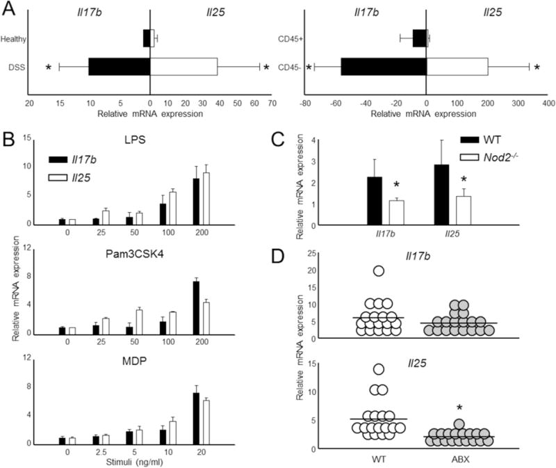
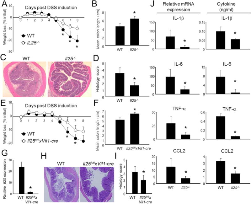
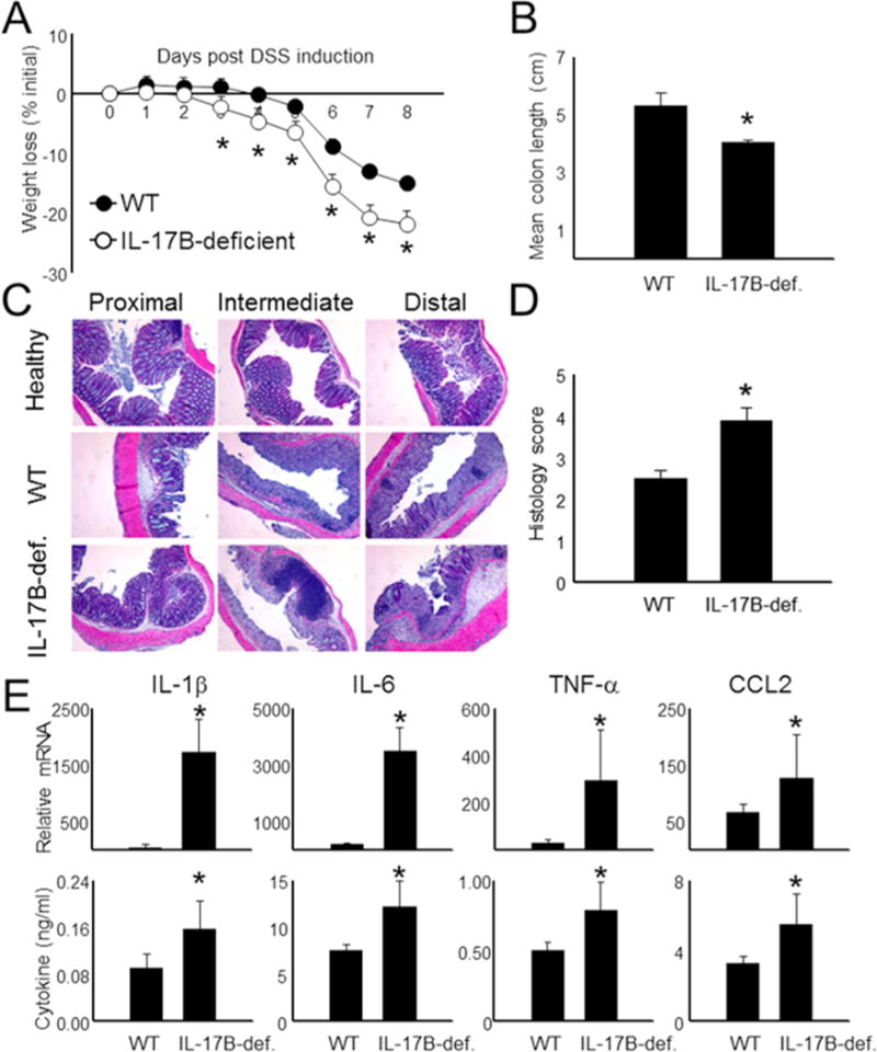
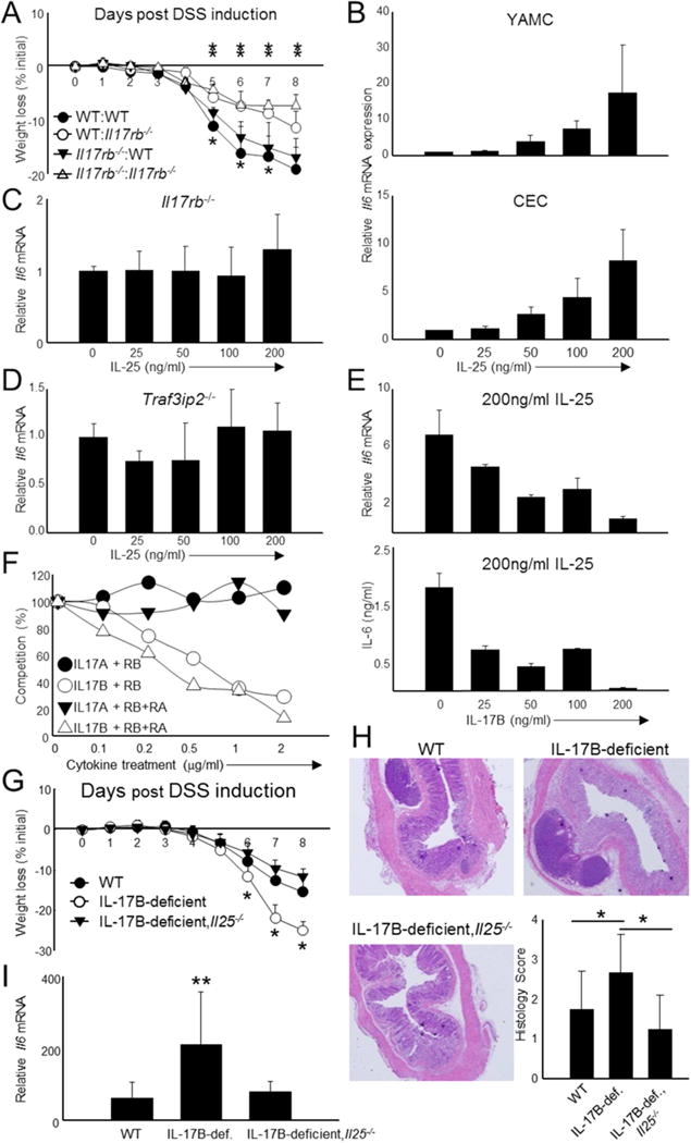
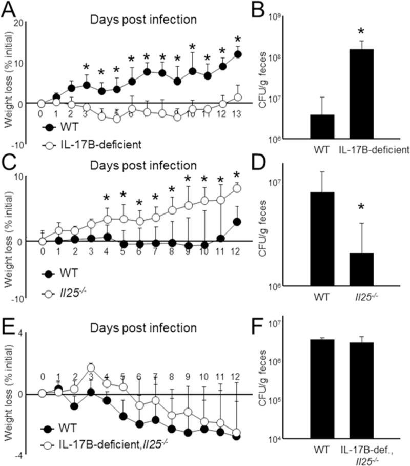
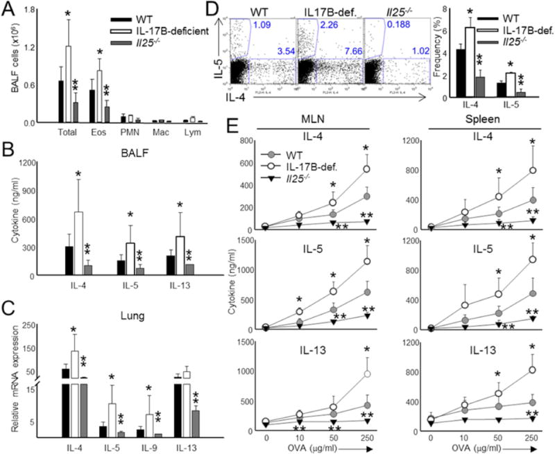
The interleukin-17 (IL-17) family of cytokines has emerged as a critical player in inflammatory diseases. Among them, IL-25 has been shown to be important in allergic inflammation and protection against parasitic infection. Here we have demonstrated that IL-17B, a poorly understood cytokine, functions to inhibit IL-25-driven inflammation. IL-17B and IL-25, both binding to the interleukin-17 receptor B (IL-17RB), were upregulated in their expression after acute colonic inflammation. Individual inhibition of these cytokines revealed opposing functions in colon inflammation: IL-25 was pathogenic but IL-17B was protective. Similarly opposing phenotypes were observed in Citrobacter rodentium infection and allergic asthma. Moreover, IL-25 was found to promote IL-6 production from colon epithelial cells, which was inhibited by IL-17B. Therefore, our data demonstrate that IL-17B is an anti-inflammatory cytokine in the IL-17 family.


Conflict of interest statement

CONFLICT OF INTEREST

The authors declare no competing financial interests.

Figures

Figure 1
Figure 1. Colon epithelial cells produce IL-17B and IL-25 in acute colitis
(A) Colon samples were derived from healthy controls or from animals following 8 d DSS-induced colitis (left). DSS samples were further processed and separated into leukocyte (“CD45+”) and epithelial cell (“CD45”) fractions (right). The relative expression of Il17b and Il25 were then measured by qPCR. n = 5 mice per group. (B) Primary colon epithelial cells derived from healthy mice were activated through TLR (LPS, Pam3CSK4) and NOD2 (MDP) pathways for 6 h. Il17b and Il25 mRNA expression was assessed by real-time RT-PCR. (C) Total colon tissue was isolated from healthy WT and NOD2-deficient animals and then assessed for Il17b and Il25 expression by qPCR. n = 5 mice per group. (D) C57BL/6 animals were left untreated (normal) or administered an antibiotic regiment (ABX) for 4 wk to deplete microbiota. Total colon tissue was then obtained and the expression of Il17b and Il25 was measured by qPCR. n = 18 mice per group. All gene values were normalized to the expression of the house keeping gene Actb. Data are presented as mean + SD and are representative of at least 3 independent experiments. * Student’s t test; p < 0.05. See also Figure S1.
Figure 2
Figure 2. IL-25-deficient mice are protected from DSS-induced colitis
(A) Acute colitis was induced in WT and Il25−/− mice by the addition of DSS in drinking water (n = 5 – 6 per group). Weight loss during colitis progression is shown. (B) Mice with colitis were euthanized on day 8 and colon length was measured from individual mice and data was combined. (C) Representative H & E histology from intermediate colon sections derived from the mice in (A). (D) Combined histological scores of all mice and all sections (proximal, intermediate, and distal) in (A). (E) Weight loss of WT and Il25fl/flxVi1l-cre mice throughout the course of DSS-induced colitis. n = 5-6 mice per group. (F) Combined individual colon lengths from the mice in (E). (G) Combined expression of total colon IL-25 mRNA from each mouse in (E). (H) Representative H & E histology from middle colon sections from mice in (E). (I) Combined histological scores from all colon sections of DSS mice presented in (E). (J) Total colon tissue was isolated from mice 8 d following the induction of DSS colitis and equivalent biopsy sections were taken for inflammatory mRNA analysis (qPCR) or were cultured in media overnight for cytokine analysis in supernatants (ELISA). The data from individual mice were combined for each group. All gene values were normalized to the expression of Actb. Data from individual mice are presented as mean + SD and are representative of at least 3 independent experiments. *Student’s t test; p < 0.05 for comparisons between WT control and Il25−/− groups.
Figure 3
Figure 3. IL-17B-deficient mice exhibit exacerbated colitis
(A) Weight loss (% of starting weight) was assessed daily in WT and IL-17B-deficient mice following DSS administration. n = 5-6 mice per group. Colons were isolated from individual mice following sacrifice at d 8 and then measured and combined for each group (B). (C + D) Colon tissue derived from the mice in (A) were sectioned and then stained with H&E to measure infiltration and inflammation in individual mice following DSS onset at d 8. (E) Total colon tissue was divided into similar sections and then combined for each mouse. mRNA was then isolated and gene values were measured by qPCR using the expression of Actb as a reference. Supernatants from the colon were also assayed for cytokine expression by ELISA. Data are presented as mean + SD and are representative of at least 3 independent experiments. *Student’s t test; p < 0.05 in comparisons of IL-17B-deficient to WT controls. See also Figure S2.
Figure 4
Figure 4. IL-17B inhibits IL-25-mediated IL-6 production in colon epithelial cells
(A) Bone marrow chimeric mice were generated using WT and Il17rb−/ animals as donors and recipients. The first portion of the legend represents hematopoietic cell origin (WT or Il17rb−/−) while the second represents non-hematopoietic cell origin. (B) IL-6 expression was measured by qPCR following 6 h stimulation with recombinant IL-25 in a colon epithelial cell line (YAMC) and primary colon epithelial cells (CEC). (C+D) IL-6 mRNA expression was similarly measured from CEC derived from IL-17RB- (C) and Act1 (Traf3ip2−/−) - (D) deficient mice. (E) IL-6 was examined in CEC following fixed IL-25 stimulation (200ng/ml) followed by simultaneous treatment of increasing IL-17B concentrations. IL-6 was measured by qPCR (6 h stimulation, top) and ELISA (24 h stimulation, bottom). (F) Summary of competition experiments measuring the ability of increasing concentrations of IL-17B or IL-17A to displace IL-25-hIg bound to murine IL-17RB (RB) or IL-17RB and IL-17RA (RB+RA). Competition (%) is the percentage of IL-25 displacement at each IL-17B or IL-17 concentration compared those treated with only IL-25. (G) Combined weight loss data from WT, IL-17B-deficient, and IL-17B and IL-17E double deficient (IL-17B-deficient,Il25−/−) mice following DSS administration. n = 7-8 mice per group. (H) Representative H & E histology and combined histological scores of all colon sections from the mice shown in (G). (I) Combined IL-6 mRNA expression from total colon samples derived from the mice in (G). Data are presented as mean + SD and are representative of 2 independent experiments (A) and at least 3 independent experiments (B-I). mRNA data was obtained by examining relative gene expression to Actb. *Student’s t test; *p < 0.05 for daily comparisons of WT:WT mice to Il17rb−/−: Il17rb−/− mice and **p < 0.05 for daily comparisons of WT: Il17rb−/− mice to Il17rb−/−: Il17rb−/− mice (A); *p < 0.05 for daily comparisons of IL-17B-deficient,Il25−/− mice to WT (G); **p < 0.05 for comparison of IL-17B-deficient,Il25−/− mice to IL-17B-deficient and to WT (G-I). See also Figure S3.
Figure 5
Figure 5. IL-17B is protective against Citrobacter rodentium infection
(A) WT and IL-17B-deficient, WT and IL-25KO (C), or WT and IL-17B-deficient,Il25−/− mice (E) mice were orally infected with 2 × 10⁹ CFU of Citrobacter rodentium and weighed daily for 12-13 d. n = 7-18 mice per group. (B-F) After harvest, feces were collected, homogenized, and then examined for Citrobacter rodentium growth on MacConkey agar after overnight incubation. Data are presented as mean + SD and are representative of at least 3 independent experiments. *Student’s t test; p< 0.05. See also Figure S4.
Figure 6
Figure 6. IL-17B and IL-25 have opposing roles during allergic asthma
(A) Total cell counts in the BAL fluid of WT, IL-17B- and IL-25-deficient animals sensitized with OVA in alum at day 0 and 14, followed by intranasal challenge with OVA at day 14, 25, and 26. (B) BALF derived from the animals in (A) was assayed for the presence of IL-4, IL-5, and IL-13 by ELISA. (C) Combined mRNA analysis of Th2 and Th9 cell-associated genes from total lung samples derived from the mice in (A). (D) Representative (left) and combined analysis (right) of Th2 cell frequencies in the BALF of OVA and alum sensitized animals. T cells isolated from the BALF were restimulated with 50μg/ml OVA prior to intracellular cytokine staining for IL-4 and IL-5. (E) Analysis of splenic and mediastinal lymph node Th2 compartments following 3 d restimulation with increasing doses of OVA. IL-4, IL-5, and IL-13 protein was measured from the supernatants by ELISA. Data are presented as mean of data obtained from individual mice + SD and are representative of at least 3 independent experiments. *Student’s t test; *p < 0.05 for comparisons of WT to IL-17B-deficient and **p < 0.05 for comparisons of WT to Il25−/−. See also Figure S5.

Comment in

References

    1. Alex P, Zachos NC, Nguyen T, Gonzales L, Chen TE, Conklin LS, Centola M, Li X. Distinct cytokine patterns identified from multiplex profiles of murine DSS and TNBS-induced colitis. Inflammatory bowel diseases. 2009;15:341–352. - PMC - PubMed
    1. Angkasekwinai P, Chang SH, Thapa M, Watarai H, Dong C. Regulation of IL-9 expression by IL-25 signaling. Nature immunology. 2010;11:250–256. - PMC - PubMed
    1. Angkasekwinai P, Park H, Wang YH, Wang YH, Chang SH, Corry DB, Liu YJ, Zhu Z, Dong C. Interleukin 25 promotes the initiation of proallergic type 2 responses. The Journal of experimental medicine. 2007;204:1509–1517. - PMC - PubMed
    1. Atreya R, Mudter J, Finotto S, Mullberg J, Jostock T, Wirtz S, Schutz M, Bartsch B, Holtmann M, Becker C, et al. Blockade of interleukin 6 trans signaling suppresses T-cell resistance against apoptosis in chronic intestinal inflammation: evidence in crohn disease and experimental colitis in vivo. Nature medicine. 2000;6:583–588. - PubMed
    1. Buning C, Genschel J, Weltrich R, Lochs H, Schmidt H. The interleukin-25 gene located in the inflammatory bowel disease (IBD) 4 region: no association with inflammatory bowel disease. European journal of immunogenetics: official journal of the British Society for Histocompatibility and Immunogenetics. 2003;30:329–333. - PubMed

Publication types

MeSH terms